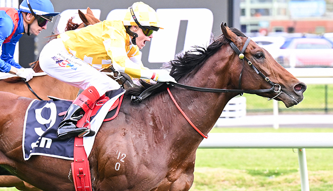
Cox Plate favourite Sir Delius has been ruled out of next week’s Group 1 and the Melbourne Cup after failing independent specialist scans.
Racing Victoria announced on Friday afternoon that the Gai Waterhouse and Adrian Bott-trained superstar and $2.80 Cox Plate favourite returned scans that showed it was currently at heightened risk of injury.
The first scan, a CT scan on the distal limbs, was conducted at University of Melbourne Equine Centre.
CT scan results are reviewed by an independent panel of diagnostic imaging specialists which comprises representatives from Australia, UK and USA.
That panel then relayed their findings to Racing Victoria.
Under the enhanced protocols in 2025, Sir Delius’ connections were then given the chance to send the star for a PET scan.
This was the first time that connections have been afforded the opportunity to utilise the state-of-the-art PET technology as part of vet protocols for the Melbourne Cup.
A PET scan shows activity of bone or soft tissue lesions at the molecular level in 3D imagery.
That scan was conducted on Thursday with the results shared with the same independent panel.
“Having reviewed the PET scan results alongside the CT scan results, the panel members have advised RV Veterinary Services that they remain of the view that Sir Delius is currently at heightened risk of injury,” an RV release read.
“Following advice from RV Veterinary Services in relation to the specialist opinions from the imaging panel, RV Stewards have stood down Sir Delius from competing in the remainder of the 2025 Spring Racing Carnival. The Stewards have appraised the connections of the key information that they relied upon in making their decision.”
Racing.com understands that Racing Victoria remained in constant contact with the Waterhouse and Bott stable throughout the decision-making process.
All horses wanting to compete in this year’s Melbourne Cup must be presented for a standing CT scan of their distal limbs by no later than Thursday, 30 October.
WHAT IS A CT SCAN?
Computed Tomography (CT) – The standing CT scanner allows efficient 3D imaging of the lower limb and identification of otherwise undetected bone damage. It is essentially cross-sectional radiographs and provides excellent, high-detailed images of bone. The quality and contrast of images created by CT is far superior to standard x-ray. It requires a mild sedative and scanning takes less than 30 minutes. In Australia, there is a standing CT scanner at the University of Melbourne Equine Centre at Werribee and another in Sydney.
WHAT IS A PET SCAN?
PET is a unique medical imaging procedure that shows activity of bone or soft tissue lesions at the molecular level in 3D imagery. PET requires a small amount of radioactive material to be injected into the horse 30 minutes before its scan which takes approximately 30 minutes and can be conducted whilst the horse is standing. The only PET scanner in Australia is located at the University of Melbourne Equine Centre at Werribee. Originating in the USA, the technology was trialled successfully during the 2024 Spring Racing Carnival before being adopted into practice in 2025.
WHO IS ON THE PANEL?
The five members of the independent imaging review panel are as follows:
Dr. Bruce Bladon, Specialist in Equine Surgery and Hospital Director at Donnington Grove Veterinary Group in England, UK;
Dr. Mathieu Spriet, Specialist in Equine Diagnostic Imaging, and Professor at the UC Davis School of Veterinary Medicine in California, USA;
Professor Peter Muir, Specialist in Veterinary Surgery and Co-Director of the Comparative Orthopaedic and Genetics Research Laboratory at the University of Wisconsin-Madison, USA;
Professor Chris Whitton, Specialist in Equine Surgery and long-time Professor of Equine Medicine and Surgery at the University of Melbourne, AUS; and
Dr. Alex Young, Specialist in Equine Diagnostic Imaging and Director of Alex Young Specialist Veterinary Imaging in Queensland, AUS.

