
But getting the healthy cells into the brain is difficult because the body tightly restricts access to the central nervous system. Other possible complications of a bodywide transplant include the elimination of donor cells by any remaining immune cells or, conversely, the life-threatening graft-versus-host disease.
Research from Wernig’s laboratory in 2022 showed it’s possible to achieve 90% engraftment of donor cells by wiping out the recipient animal’s immune system and treating them with a drug to kill off existing microglia, giving the donated hematopoietic stem cells a competitive growth advantage. But the technique still required toxic preconditioning and relied on genetically identical donor cells to prevent graft-versus-host disease.
“A hematopoietic stem cell transplant is a rough procedure to go through,” Wernig said. “It’s not something you want to do to your patients unless there’s no other option.”
A targeted transplant procedure
In the current study, Mader and Wernig tested whether they could develop a brain-specific transplant procedure that would avoid the toxic preconditioning and bodywide effects of a hematopoietic stem cell transplant. To that end, they coupled the microglia-depleting drug treatment with irradiation of the brain to create space for the new cells to occupy. They then injected microglia precursor cells — a more specialized subset of hematopoietic stem cells — from a non-genetically matched donor animal into the brain. Finally, they administered two drugs to block the activation of immune cells from elsewhere in the body that would otherwise kill the unmatched donated cells.
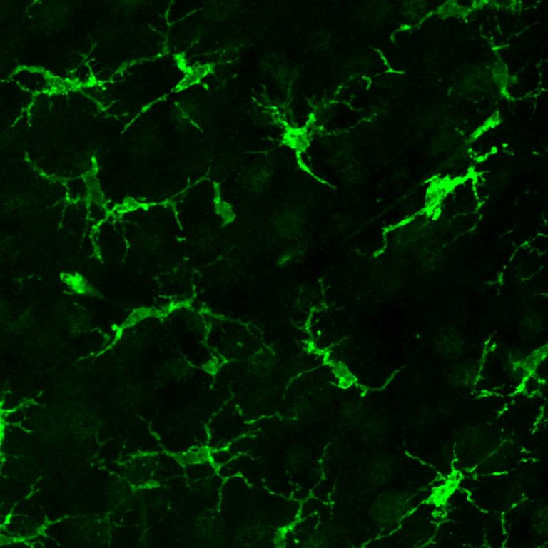
This delicate tango of steps resulted in efficient engraftment of the donated cells, which nestled into the brain and developed into microglia without migrating to the rest of the body or being attacked by the recipient animal’s immune system.
The cells’ engraftment was lasting: More than 85% of the microglial cells in the brain were derived from the donated cells eight months after transplantation. Untreated mice with a version of Sandhoff disease lived a median of 135 days and no animal lived beyond 155 days. In contrast, five of the animals treated with the brain-specific microglia transplantation therapy lived up to 250 days, when the experiment was terminated.
Although the long-lived treated mice eventually developed hind leg paralysis, they displayed normal exploration behaviors in a large open pen and greater muscle strength and coordination than control animals.
A closer investigation of the relationship between the donated microglia and their neuronal neighbors revealed something intriguing: The missing lysosomal enzyme now being made by the microglia could also be found in the animals’ native neurons. Although the reason is not yet known, Wernig and Mader suspect the microglia are packaging and secreting the enzyme into the space between cells, where it is imported into the neurons.
“This could be an important, unrecognized role for microglia: to supply lysosomal factors to the environment including neurons,” Wernig said.
The researchers are optimistic their approach could be translated to humans because each individual step — irradiation, administering drugs used to wipe out existing microglia and applying drugs to prevent immune attack of the donated cells — is already used to treat other conditions.
“We’ve solved three big problems with this study,” Wernig said. “We achieved efficient brain-restricted transplantation without systemic toxic preconditioning, we were able to use non-genetically matched cells that don’t require genetic engineering to make the missing lysosomal enzyme, and we avoided immune rejection and graft-versus-host disease. We’re very happy.”
The researchers also believe the therapy could be widely applicable.
“It’s possible that these lysosomal storage diseases are just an accelerated version of much more common neurodegenerative diseases like Alzheimer’s or Parkinson’s,” Wernig said. “If so, this therapy could be very relevant not just for a small subset of children, but for many, many more people.”
The study was funded by the California Institute for Regenerative Medicine, the German Research Foundation, the New York Stem Cell Foundation, the Robert J. Kleberg Jr. and Helen C. Kleberg Foundation, and the Wu Tsai Neurosciences Institute.