
Our main finding was no significant H3K27me3 differences in any TET1 promoter region between endometriosis and controls, regardless of ChIP efficiency. This contrasts with our hypothesis, suggesting a more complex H3K27me3–TET1 relationship in endometriosis. The absence of differences across TET1 promoter regions, even after stratifying by fertility and cycle phase, suggests H3K27me3 changes may not drive endometriosis-related infertility.
However, region 2 showed higher H3K27me3 in endometriosis (p = 0.04) in the good ChIP efficiency subgroup. This highlights the importance of considering technical factors such as ChIP efficiency when interpreting epigenetic data. The region 2 difference, limited to the good efficiency subgroup, warrants further study. This region may be sensitive to technical variation or reflect a real difference masked by technical noise. The low test power increases the risk of type II error, suggesting the effect may be too small to detect in this diverse sample. This may suggest that the effect is too small to detect in this diverse sample, highlighting the need for stricter analysis and larger groups22,23.
This study included fertile endometriosis patients to explore H3K27me3 in infertility but was limited by whole-tissue analysis. Stem cell theory suggests circulating progenitors form stroma and glands at ectopic sites, creating endometriotic lesions. Noë et al. showed ectopic epithelial and stromal cells develop clonally and independently, forming distinct compartments14.
Mahajan et al. found estrogen-dependent TET1 expression in eutopic endometrium is mainly in stromal cells15. Roca et al. examined which compartment mainly drives TET expression in endometriosis-affected endometrium24. Epithelial cells expressed TET transcripts 10–30 times higher than stromal cells, while stromal fibroblasts showed significantly reduced TET mRNA vs. controls. These differences may reflect stromal cell enrichment, which largely drives altered TET expression. Thus, stromal TET expression seems key in defective TET regulation.
Future epigenetic studies should analyze specific cell compartments, not whole tissue, to avoid misinterpretation24,25,26,27. Future studies should use laser capture microdissection or cell-type-specific chromatin profiling.
ChIP-seq of decidual vs. myometrial stromal cells in mice revealed > 800 differential peaks, highlighting the need for cell-type-specific analysis28. The lack of significant overall H3K27me3 differences may have several causes.
Endometriosis-related infertility involves many pathways, and H3K27me3 changes may be just one piece of this puzzle. Other epigenetic or genetic changes may be more important. Our small sample size may have limited power to detect subtle H3K27me3 changes. Environmental, genetic, and hormonal factors affecting epigenetics also warrant consideration.
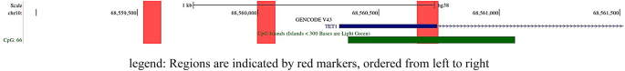
H3K27me3 and H3K9me3 are key repressive marks in promoters of silenced genes, including TET1. The balance between H3K27ac and H3K27me3 further complicates TET1 regulation. These interactions highlight the need for genome-wide methylation analysis with H3K27me3 profiling to understand endometriosis epigenetics29.
EZH2, a PRC2 methyltransferase, catalyzes H3K27 methylation30. EZH2 gain-of-function mutations raise H3K27me3 and suppress differentiation31,32. EZH2 overexpression promotes proliferation, epithelial–mesenchymal transition, and invasiveness33. Endometriotic epithelial cells show higher progesterone-regulated EZH2 than non-endometriotic cells11. Elevated EZH2 in endometriotic tissue suggests a role in pathogenesis11,34. The EZH2–PRC2–H3K27me3 axis regulates endometrial differentiation and implantation receptivity35. Inhibiting histone methyltransferase activity reduces endometriotic cell growth and invasiveness, supporting EZH2 as a therapeutic target11. We did not assess EZH2 expression. Further research should examine EZH2 expression and its link to H3K27me3 in endometriosis.
Few studies examined H3K27me3 in the TET1 promoter, mostly in cancers with low TET1, similar to endometriosis36,37. These studies suggest EZH2 may regulate TET1, warranting studies on EZH2 expression and H3K27me3 in eutopic endometrium. Other studies linked active histone marks, such as H3K4me3, to higher TET1 expression25,38. No studies examined H3K27me3 in TET1 in eutopic or ectopic endometrium in endometriosis.
H3K27me3 represses developmental genes (e.g., HOX) and COX-2, lowering PGE239,40. Because these genes are involved in endometriosis, further research into H3K27me3 is warranted. Epigenetic changes are reversible but harder to reverse as they accumulate. Epigenetic mechanisms may offer non-hormonal therapies for endometriosis. Identifying gene expression patterns in eutopic and ectopic endometrium may enable therapies targeting epigenetic regulators in key promoters.
In conclusion, we found no overall H3K27me3 differences in the TET1 promoter between endometriosis and controls, except for one significant difference in region 2 (p = 0.04) with good ChIP efficiency, confirmed after multiple testing correction. This finding should be interpreted cautiously due to the small sample size. The lack of consistent subgroup differences limits biological conclusions. The complex histone interactions, multifactorial infertility, and methodological issues highlight the need for larger cohorts, comprehensive epigenetic profiling, and optimized methods. Cell-type-specific analyses are likely to be especially informative. Such studies are essential to clarify the role of epigenetics in endometriosis and infertility.
The data are available from the corresponding author upon request.