Defensive response could help restrict tumour spread
Cancer cells have been shown to mount a rapid, energy-intensive defence when physically compressed, according to a study which has revealed a previously unrecognised survival mechanism that enables cancer cells to resist DNA damage and survive the physically constrained conditions of the body.
This response may help explain how cancer cells endure the mechanical challenges of their microenvironment, from forcing their way through tissues in order to enter blood vessels or withstand the turbulence of the circulatory system. The authors suggested that targeting this mechanism could offer a novel therapeutic strategy to contain tumour spread.
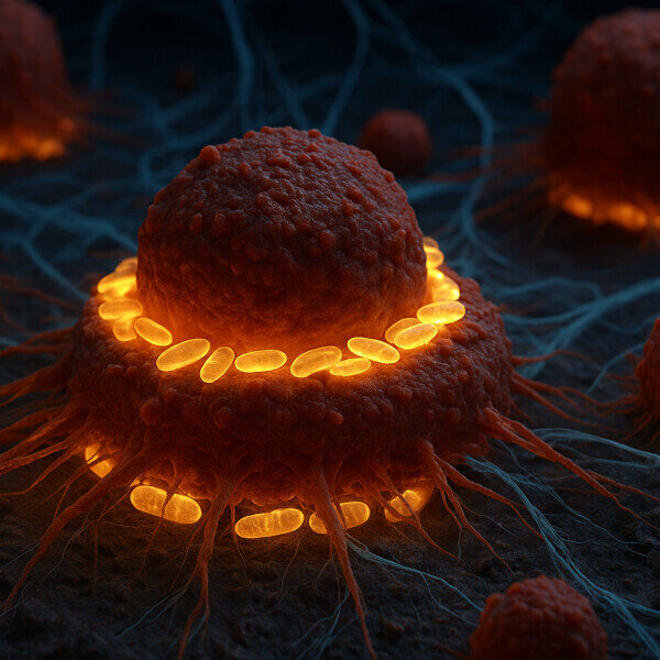
Researchers at the Centre for Genomic Regulation (CRG) in Barcelona observed the phenomenon by using a specialised microscope to compress living HeLa cells to just three microns wide. Within seconds, mitochondria migrated to the nuclear membrane and delivered a rapid influx of adenosine triphosphate (ATP) – the molecular energy currency of cells.
This response was visible in 84 percent of confined cancer cells, but almost absent in those in suspension. The team termed the structures; ‘nucleus-associated mitochondria’ – or NAMs.
“We must reconsider the role of mitochondria,” said Dr Sara Sdelci, co-corresponding author.
“They are not passive batteries but agile responders summoned [upon] when cells are physically pressed to the limit.”
To investigate NAM function, the team used a fluorescent probe to detect ATP levels inside the nucleus. The signal increased by approximately 60 percent within three seconds of compression.
“It is clear the cells adapt to strain by rewiring their metabolism,” stated Dr Fabio Pezzano, co-first author.
Further experiments indicated the ATP surge supported DNA repair. Physical compression disrupts the genome, but ATP-dependent repair mechanisms enabled cells to fix damage and resume division. Cells that failed to receive the energy boost did not divide correctly.
To test clinical relevance, the researchers analysed breast tumour biopsies from 17 patients. NAMs were found in 5.4 percent of nuclei at invasive tumour margins, compared with 1.8 percent in the tumour core.
“Seeing this signature in patient samples confirmed its broader significance,” said Dr Ritobrata Ghose, co-first author.
The study also examined how NAMs are physically stabilised. Actin filaments formed a contractile ring around the nucleus, while the endoplasmic reticulum created a mesh-like net. This scaffold physically trapped mitochondria at the nuclear envelope. When the Actin network was disrupted with latrunculin A, NAM formation collapsed and ATP delivery ceased.
If metastatic cells rely on NAM-driven ATP surges, inhibiting this scaffold could reduce invasiveness without harming healthy mitochondria.
“Mechanical stress responses are an underexplored vulnerability of cancer cells,” said Dr Verena Ruprecht, co-corresponding author.
“They may represent a promising route for future therapies,” she added.
Although the work focused on cancer, the authors emphasised that similar pressures affect other cell types. Immune cells, neurons, and embryonic cells all experience physical confinement during development and function.
“Wherever cells are under pressure, a nuclear energy boost is likely safeguarding genomic integrity,” concluded Dr Sdelci.
“It reveals a fundamental layer of regulation in cell biology and changes how we understand survival under mechanical stress.”
For further reading please visit: 10.1038/s41467-025-61787-x

