
Heart disease remains a primary cause of illness and mortality around the world. While minimally invasive procedures have improved cardiac care, the heart’s complex anatomy and constant motion require rigorous hands-on training to reduce procedural errors.
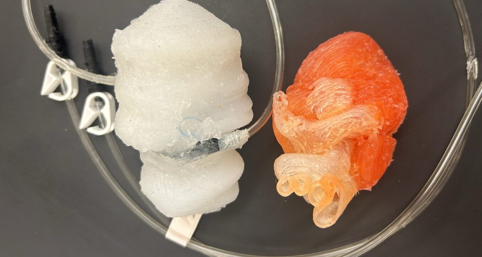
To address this issue, researchers have developed a 3D-printed dynamic heart model that mimics the complex movements of the left side of the heart. This model includes atrium, ventricle, and mitral valve, offering surgeons a realistic platform to practice patient-specific presurgical simulations.
Realistic physiological engineering
In the past, dynamic heart models have relied on animal tissues to mimic pumping mechanics. However, this approach raised significant ethical concerns and faced policy restrictions. While synthetic models exist, it has been very challenging to replicate the full dynamics of a beating heart.
This new research deploys soft material 3D-printing to accurately recreate anatomical features. To enhance structural realism, sutures connect the ventricle to the mitral valve, imitating the chordae tendineae—the “heartstrings” that support valve function. These features ensure the model behaves like real tissue during practice procedures.
Replicating cardiac contraction
A key innovation in this model is the use of McKibben actuators placed inside the heart muscle walls. These soft robotic parts contract to copy the movement of the ventricle and the natural motion of the mitral valve.
To give feedback during use, the team added custom flexible pressure sensors. These sensors track pressure changes inside the model, making it a useful tool for studying blood flow. According to the research, “the heart’s anatomical complexity and dynamics require proper hands-on training on patient-specific presurgical models to reduce procedural errors.”
Enhancing surgical outcomes
By combining soft robotics with 3D printing, this research provides a synthetic option to avoid the ethical issues related to animal testing while keeping the physical realism required for medical training. This progress is an important step toward personalized medicine, helping make surgical practice as accurate as possible to improve patient outcomes.
This model is designed to support the simulation of edge-to-edge repair, a method used to fix atrioventricular valve leakage. By practicing on a model that copies heart circulation and contraction, doctors can improve their technique for complex valve repairs before surgery.