
Researchers have identified a previously overlooked neural pathway that helps control human hand and arm movements.
The discovery shows that signals guiding voluntary hand motion travel through deeper brainstem and spinal relays, expanding how scientists understand the nervous system’s control of dexterity.
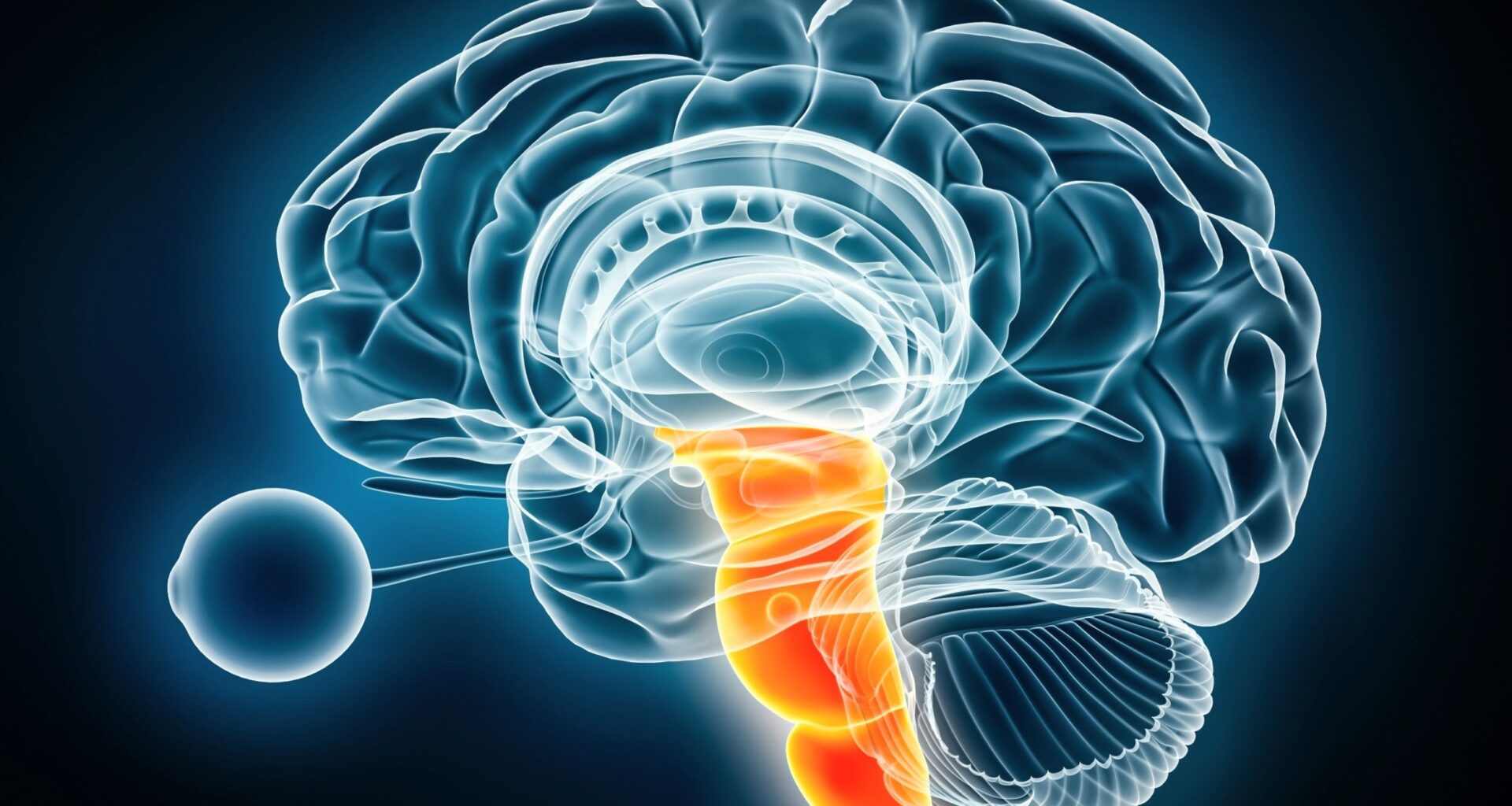
Brain scans reveal a new pathway

Activity patterns in the lower brainstem and upper spinal cord revealed a hidden relay that becomes active when hands grip or apply force.
Working at the University of California, Riverside (UCR), Dr. Shahab Vahdat, Ph.D., traced those signals through two small hubs in the brainstem while examining human and animal movement.
Those same regions remained tightly connected with motor areas of the brain during hand actions, indicating that voluntary movement flows through this relay rather than a single direct command.
Recognizing that extra pathway reframes how signals reach the hand and sets the stage for exploring how those circuits might support recovery after injury.
Activity in the brainstem
Deep in the brainstem, two spots in the medulla, the lowest part just above the spinal cord, stayed active through the gripping tasks.
Rather than merely passing commands along, those hubs seemed to sort and blend incoming signals before sending instructions onward.
“For a long time, we thought fine hand movements in humans were controlled almost entirely by the cortex,” said Vahdat.
That older control layer may explain why the hand keeps some options even when cortical motor areas fail.
The same route in other mammals
Matching activity in mice and people mattered because it showed the route was not a human one-off.
Animals learned to press a small lever, while volunteers squeezed a device at different force levels inside the scanner.
“Despite the differences between our brains, we found striking similarities in how these regions communicate,” said Vahdat.
Because evolution rarely keeps useless circuitry, that overlap makes the pathway more than a minor side route.
Circuits that activate fingers
Another surprise appeared in the upper neck, where two spinal segments took on more than a simple delivery job.
Those segments, called C3-C4, sit in the upper neck portion of the spinal cord and pass signals to lower levels.
Instead of simply transmitting signals, this relay appeared to link brainstem commands with the lower spinal circuits that activate fingers.
That extra relay helps explain how the nervous system can refine grip and force before muscles contract.
The brain doesn’t work alone
None of this removed the cortex, the brain’s outer layer responsible for conscious thought and voluntary movement, from the picture, but it did cut against the idea of solo control.
Signals still seem to start in higher motor areas, then travel through the brainstem and upper cord before cells finally activate muscles.
That layered route likely lets different regions add timing, posture, and force adjustments while the movement is already underway.
Fine finger control may still depend most on the direct route, yet the indirect path clearly contributes.
Implications for stroke patients
Stroke makes this discovery more than an anatomy story because hand weakness can persist long after people survive the initial injury.
When damage hits the corticospinal tract, the main highway from cortex to spinal cord, fine hand control often drops hardest.
Older evidence still showed that damage along that highway tracks closely with stroke-related motor severity.
A surviving relay below the injury will not solve everything, but it could give therapists another workable target.
Targets for recovery
Because the pathway remains connected to movement, it gives clinicians more places to try nudging the system.
That could include neuromodulation, controlled stimulation that changes nerve activity, aimed at circuits the stroke did not erase.
“These pathways give us additional targets to explore,” said Vahdat, pointing to circuits that may still be reachable after cortical damage.
Even so, turning a mapped circuit into therapy will require proof that stimulation improves real hand use, not only scans.
The pathway was hard to detect
Scientists missed this route for so long partly because the relevant tissue is small, deep, and hard to image during movement.
The team used functional MRI, a scan that tracks blood-flow changes linked to neural activity, across brainstem and spinal cord.
Reading both the brainstem and spinal cord at once mattered because signals can look unrelated when each piece gets measured alone.
Better imaging did not invent the pathway, but it finally let researchers watch the relay in action.
New map of hand movement control
Early findings like these still come with boundaries, even when the evidence already looks convincing.
Brain activity can reveal coordinated traffic, but it cannot by itself prove which region issued the decisive command.
Seventeen healthy volunteers and fifteen trained mice provided strong early evidence, yet studies in patients recovering from stroke still need to follow.
The next step is plain enough: show that this hidden relay can be trained or stimulated after injury.
Hand movement now looks less like a single order and more like a chain that runs through older neural machinery.
That deeper map widens the search for recovery, while also reminding researchers that regaining dexterity may depend on several surviving links.
The study is published in the journal Proceedings of the National Academy of Sciences.
—–
Like what you read? Subscribe to our newsletter for engaging articles, exclusive content, and the latest updates.
Check us out on EarthSnap, a free app brought to you by Eric Ralls and Earth.com.
—–