
A new brain imaging system, Connectome 2.0, is far outperforming traditional MRIs in imaging microscopic brain structure disruptions resulting from neurological and neuropsychiatric disorders, potentially leading to the first comprehensive brain maps and new individualized treatments, according to a new paper on the developments.
The new MRI scanner, supported through the National Institute of Health’s (NIH) Brain Research through Advancing Innovative Neurotechnologies (BRAIN) Initiative, makes a significant leap in neuroscience by bridging brain regions to map the connection structure of nervous system nodes, termed the “connectome,” for the first time. Notably, the device operates noninvasively, enabling scan resolutions previously only possible through post-mortem dissections.
Innovations in Brain Scanning
“This research is a transformative leap in brain imaging – pushing the boundaries of what we can see and understand about the living human brain at a cellular level,” John Ngai, Ph.D., the director of the BRAIN Initiative, commented on the new research. “The new scanner lays essential groundwork for the BRAIN CONNECTS program’s ultimate goal of developing a wiring diagram for the human brain.”
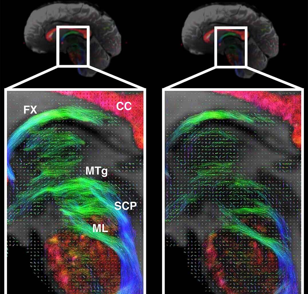
MRI machines function by using the random thermal motion of water to probe brain tissue. Advances made with Connectome 2.0 serve to push the signal-to-noise ratio higher, leading to the clearest images ever produced of extremely small structures in the brain, vastly improving from traditional MRIs and even from Connectome 1.0’s advances. Researchers accomplished this feat by developing a device that fits directly on the human head, while containing many more channels than conventional MRI machines.
The imaging is so precise that neuroscientists will be able to map the brain structure almost down to a single micron, clearly imaging individual fibers and cell structures. This will lead to a new level of granularity in the data, exploring the relationship of minute cellular and connective alterations to human experiences in cognition, behavior, and disease.
Real World MRI Results
Refinements to the Connectome’s physical structure from the 1.0 version enabled a wider range of human body sizes to utilize the device. Tests on research volunteers demonstrated that the device was safe and effective, providing clarity previously achieved only in animal and post-mortem studies. Comparisons of the results between volunteers allowed for precise identification of subtle differences in axon diameters and cell sizes. Axon diameters provide essential information on conduction velocity, while variations in cell size are indicators of neurodegeneration and aging.
“Our goal was to build an imaging platform that could truly span scales – from cells to circuits,” said senior author Susie Huang, M.D., Ph.D., of the Department of Radiology at Mass General Hospital. “It provides researchers and clinicians with a powerful new tool to study brain architecture in health and disease, in real time.”
Impact on Neuroscience
The human brain remains a mysterious and incredibly complicated structure to neuroscientists. Connectome 2.0 is a step toward removing some of that mystery by mapping the complete wiring of the human brain for the first time. By capturing extremely high-resolution images, the new MRI enables a better understanding of the brain’s smallest scales, comprising individual cells and their connections, thereby facilitating a deeper comprehension of how larger brain systems, such as whole regions and connecting circuits, function and drive human thought.
In addition to mapping, Connectome 2.0 could advance treatment options. Theoretically, the device could lead to not only noninvasive scanning but also noninvasive stimulation of brain circuits, allowing for individually designed treatments for brain disorders based on a person’s unique brain mapping.
The paper “Ultra-high Gradient Connectomics and Microstructure MRI Scanner for Imaging of Human Brain Circuits Across Scales” appeared on July 16, 2025, in Nature Biomedical Engineering.
Ryan Whalen covers science and technology for The Debrief. He holds an MA in History and a Master of Library and Information Science with a certificate in Data Science. He can be contacted at ryan@thedebrief.org, and follow him on Twitter @mdntwvlf.