
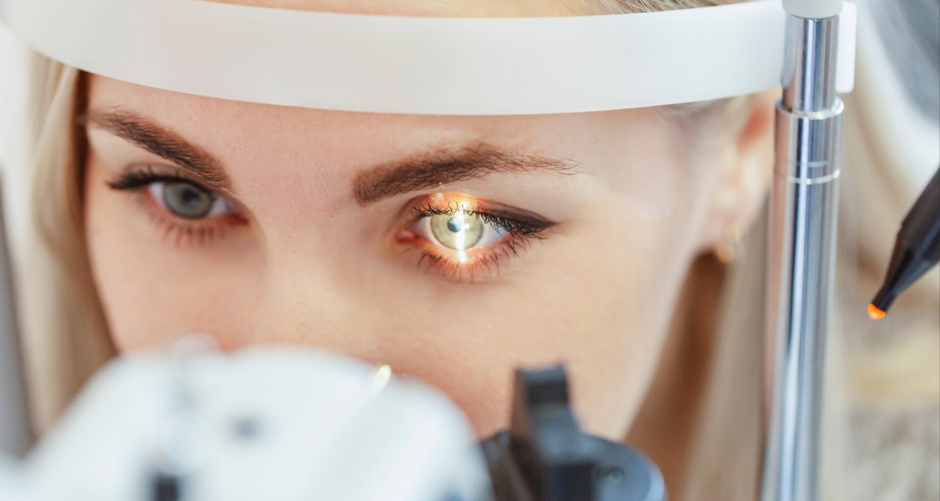
The back of the eye is constantly producing waste, shifting fluid and clearing away cellular debris. For more than a century, scientists have struggled to explain exactly where all of it goes.
That question matters because some of the leading causes of blindness involve material building up near the retina, the delicate tissue responsible for vision.

Yet researchers had never found the lymphatic drainage system that normally handles this kind of cleanup elsewhere in the body.
Now, scientists may have found the missing route.
In mice, a tracer injected behind the retina traveled out of the eye and reached lymph nodes in the neck within just 20 minutes.
The discovery reveals a previously unknown drainage pathway that could reshape researchers’ understanding of glaucoma, macular degeneration and other serious eye diseases.
Scientists thought nothing was there
Nearly every organ carries lymphatic vessels, the thin-walled channels that collect leftover fluid, haul away waste proteins and deliver immune cells to lymph nodes. The eye was the exception.
That assumption began to crack in 2009, when a paper identified lymphatic vessels in the ciliary body at the front of the human eye. Its authors named the route the uveolymphatic pathway.
Two of them have spent the years since asking whether the back of the eye had something comparable.
Dr. Neeru Gupta, professor and head of ophthalmology and visual sciences at the University of British Columbia (UBC), led the new work.
She collaborated with Dr. Yeni Yücel, professor and director of ophthalmic pathology at the University of Toronto (U of T).
The front of the eye was the easier place to look. Sight happens at the back, and so does most blindness.
“The retina is responsible for vision and it’s also where many of the most serious vision-loss diseases occur,” said Yücel.
Watching the fluid escape
Working with anesthetized mice, the team injected tracers made from a common blood protein into the suprachoroidal space.
This thin gap sits between the eye’s tough outer wall and the vessel-packed layer beneath the retina.
One tracer showed up on MRI scans. A second one glowed under near-infrared light, which let the researchers watch fluid move inside a living animal instead of reconstructing its path from tissue slices afterward.
The fluid did not seep out in every direction. It ran toward the nose-facing side of the eye socket, taking the same route each time.
Twenty minutes after injection, imaging of the head and neck picked up the tracer in a lymph node on the same side as the treated eye.
The injected side carried far more signal than the other. Fluid spreading at random would not do that.
Vessels hiding behind the eye
The choroid is a dense mat of blood vessels pressed between the retina and the white wall of the eye. It supplies most of the oxygen the retina burns through.
Earlier searches for lymphatic vessels in that layer came up empty, and the field largely stopped looking.
Under a confocal microscope, choroidal vessels in these mice carried three proteins that appear together on lymphatic tissue and not on ordinary blood vessels. The injected tracer sat inside those same vessels.
That combination is what turns a suspicion into a pathway. Until this study, no one had followed fluid from behind the retina, through vessels in the choroid, and out to the lymphatic system as one continuous route.
The team called it the POLO pathway, short for posterior ocular lymphatic outflow.
“We were surprised to see such a direct route for fluid to leave the eye and connect with the lymphatic system,” said Yücel.
The retina needs constant cleaning
“The retina is one of the most metabolically active parts of the body, constantly generating byproducts that need to be cleared,” said Gupta.
Light-sensing cells shed the worn tips of their structures every day, and the layer of cells beneath them swallows the discards.
Proteins leak out of vessels. Immune cells come and go and leave debris behind. All of it has to end up somewhere.
A 2022 review of the field described how poorly mapped that territory remained. Scientists had extensively studied drainage at the front of the eye, while the back remained largely guesswork.
The waste that builds up in glaucoma and macular degeneration has always looked like a failure of cleanup. Nobody could say what was failing because the cleanup system had not been found.
A route that drugs could use
Eye clinics already inject drugs into the suprachoroidal space, so the entry point used here is not unusual. What the results suggest is that whatever goes in does not simply pool and wait to be absorbed.
Gupta and Yücel showed in an earlier study that a common glaucoma eye drop increased lymphatic drainage from the eye in mice. Whether the newly mapped route responds the same way has not been tested.
“This gives us a whole new framework for understanding these diseases and a potential target for therapeutics,” said Gupta.
All of this was done in mice, and how closely the human eye follows the same design remains open.
The 2009 finding at the front of the eye was confirmed in human tissue, which gives the team reason to think the search is worth making.
Rethinking how the eye works
The back of the eye has a drainage route, and it appears to end in the lymphatic system.
Fluid, proteins and inflammatory material leaving the space behind the retina reach lymph nodes in the neck within minutes.
They travel along vessels that researchers spent a century assuming were absent.
That gives researchers something concrete to test in diseases that take sight away. If clearance along this route slows with age or illness, it becomes something to measure rather than something to infer from swollen tissue.
It also changes how researchers think about drugs delivered to the back of the eye. Anything placed there likely has an exit, which affects how long it lingers and where it travels.
“This is a foundational discovery that shows the eye is not as closed a system as we previously thought,” said Gupta.
The study is published in journal Translational Vision Science & Technology.
—–
Like what you read? Subscribe to our newsletter for engaging articles, exclusive content, and the latest updates.
Check us out on EarthSnap, a free app brought to you by Eric Ralls and Earth.com.
—–