
 Credit: Steven Gschmeissner/Getty Images
Adaption of an imaging tool routinely used by ophthalmologists can provide detailed images of the inner ear which could lead to earlier and more precise diagnoses of sensorineural hearing loss, according to researchers at the Keck School of Medicine at University of Southern California. The proof-of-concept study, published in Science Translational Medicine, showed how the noninvasive technology called optical coherence tomography (OCT) could be used to visualize the cellular and subcellular structures of the cochlea.
“These findings are exciting because hearing loss can happen very suddenly, and we often don’t know why, said senior author John Oghalai, MD, professor in the department of otolaryngology–head and neck surgery at the Keck School of Medicine. “OCT offers a way to explore the underlying cause and potentially guide treatment.”
In its ophthalmology application, OCT uses light waves to take cross-sectional images of the retina. It measures the reflection of light from different tissue layers to construct detailed images, allowing for the early diagnosis of retinal diseases. Researchers in this study applied similar principles to cochlear tissue by engineering OCT systems to account for the anatomy and optical properties of the inner ear.
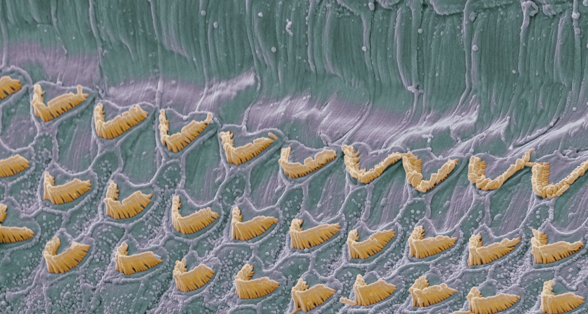
Specifically, the researchers modified OCT to capture images of the cochlea’s sensory epithelium. This includes stereocilia, the hair in the cochlea that translated sound vibrations into neural signals. Current diagnostic tools for hearing loss, such as audiograms and otoacoustic emissions testing, are indirect and lack spatial resolution of the cochlear structures. Or, as the researchers wrote, “Current clinical tools do not provide the sensitivity or resolution needed to identify sensory cell damage in vivo.”
The researchers pursued their line of inquiry based on earlier studies seeking to find if OCT could be adapted for inner ear imaging. “Recent animal studies using OCT have suggested that structural changes in the organ of Corti can be detected before measurable hearing loss occurs,” the researchers wrote, noting that these findings prompted the them to investigate its potential utility in human temporal bone samples.
In the current study, researchers used modified spectral-domain OCT to image freshly harvested human cochlear tissue. They examined samples from individuals with known histories of normal and impaired hearing. The imaging allowed the team to differentiate between intact and damaged regions of the organ of Corti based on reflectivity patterns. They found that “OCT imaging revealed reduced contrast in areas of presumed sensory cell loss and maintained structural integrity in regions associated with normal hearing.”
The findings could have implications for earlier diagnosis of sensorineural hearing loss, which affects millions of people worldwide. By identifying cellular damage before hearing loss becomes measurable through current tests, clinicians may have the opportunity to intervene. It could also guide development of targeted therapies. With the potential to visualize treatment effects on cochlear structures, OCT might one day serve as both a diagnostic and monitoring tool.
Based on success of this new study, the team will now seek to adapt the imaging system for in vivo use, either through the ear canal or during cochlear implant surgeries. Clinical trials are also being planned to test the feasibility of using OCT in patients with varying degrees of hearing loss.