
Ultrafast lasers are essential to a multitude of science and industrial applications ranging from semiconductor lithography to fundamental studies of atoms and electrons. Their pulses are often generated through using nonlinear interactions of pump lasers with a gain medium that can result in pulses with durations on the attosecond (10-18) timescale, or one billionth of a billionth of a second—timescales needed for studying the fastest processes in nature.
Our recent paper, published in the journal Nature, reports the observation of two hallmark nonlinear optical phenomena: Rabi cycling and filamentation, not in optical light but at hard x-rays with angstrom (0.1-nm) wavelengths. These effects, known within microwave to visible light laser systems, have now been observed within the x-ray regime during stimulated emission of copper (Cu) and manganese (Mn) inner-shell transitions pumped with highly intense x-ray free-electron laser (XFEL) pulses at the Linac Coherent Light Source (LCLS) at SLAC National Accelerator Laboratory in the U.S. and the SPring-8 angstrom Compact Free Electron Laser (SACLA) in Japan. The researchers find that these effects can lead to the generation of x-ray pulses with durations shorter than 100 attoseconds. What was created was the first attosecond atomic x-ray laser and the shortest hard x-ray pulses to date.
This research demonstrates how traditional nonlinear methods used at optical wavelengths can be extended to 3,000x shorter x-ray wavelengths, opening possibilities for new attosecond x-ray research with atomic spatial resolution. The resulting pulses can lead to several potential applications, from quantum x-ray optics to visualizing electron motion inside molecules.
A surprise result
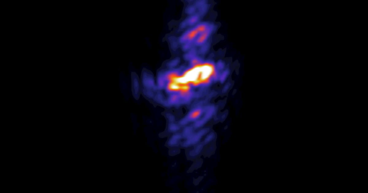
The observation of both strong lasing phenomena was not the original goal of the work with the experimental team first studying how to use stimulated x-ray emission for chemical speciation of transition metal compounds like manganese. While investigating these samples, the researchers saw multiple hotspots and random spurting in the stimulated emission, where a narrow signal was expected.
When increasing the XFEL intensity, the team started to observe additional large spectral broadening and peak splitting, much larger than possible based on their knowledge of Mn and Cu inner-shell transitions. It took them several years to identify and verify their findings. A breakthrough occurred when a recently developed three-dimensional (3D) simulation was coded to simulate the experimental conditions, rigorously showing the causes of the observed spectra.