
Researchers have discovered that pain-sensing nerve cells survive by importing mitochondria directly from nearby support cells.
Disrupting that transfer causes neurons to misfire and deteriorate, exposing a previously unrecognized mechanism behind chronic nerve pain.
Energy gifts in nerves

Inside the dorsal root ganglia – clusters of nerve cells near the spinal cord – support cells surrounded pain-sensing neurons and supplied them with extra energy.
Experts at Duke University traced that power handoff in fine detail across mouse cells, live mice, and donated human tissue.
Dr. Ru-Rong Ji showed that mitochondria, cell parts that turn oxygen into usable energy, moved into neurons from nearby cells.
With some nerve fibers stretching more than 3 feet, a fresh batch arriving locally may prevent energy shortfalls far from the cell body.
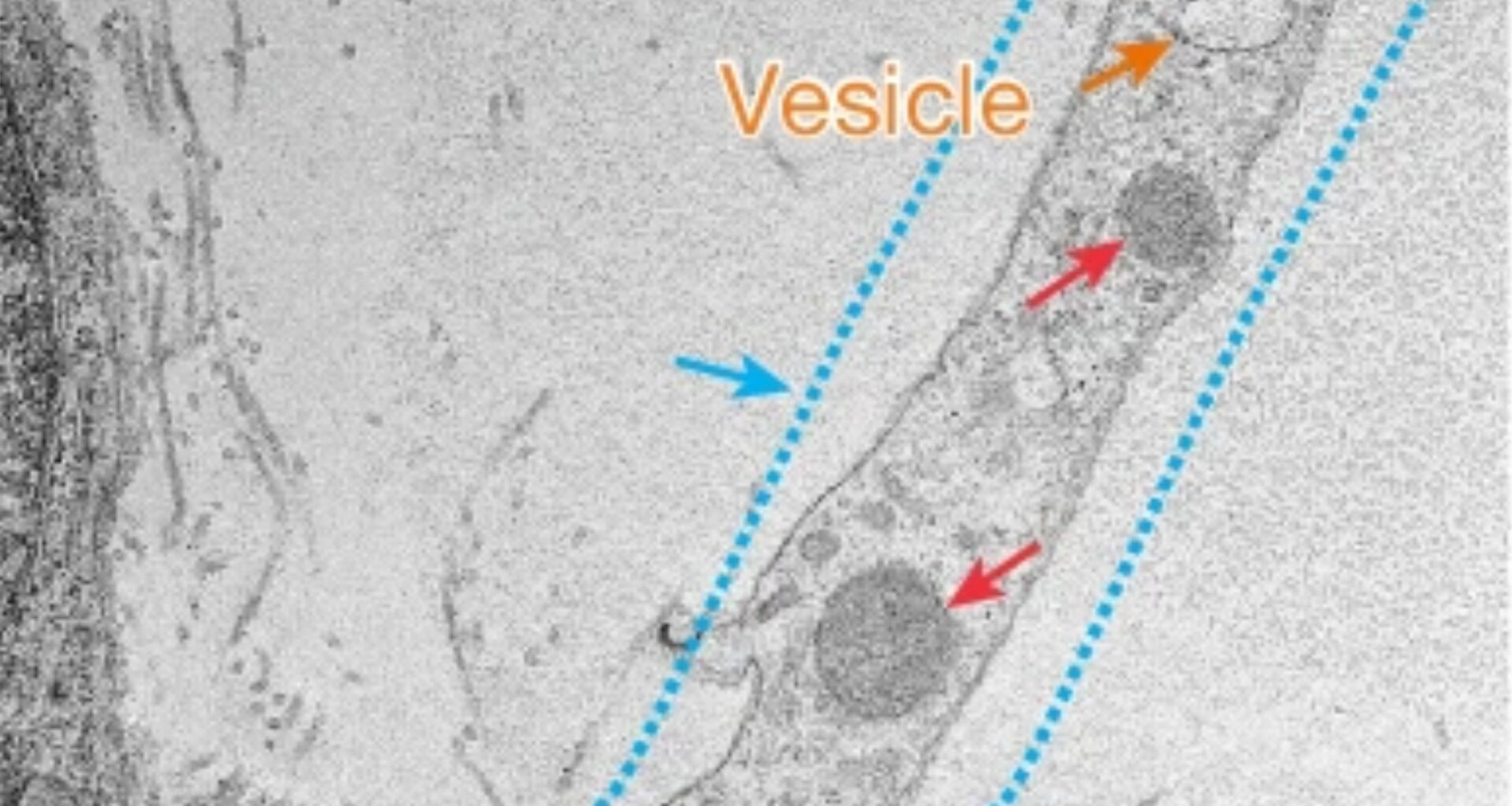
Tubes that appear and vanish
Satellite glial cells, the support cells that surround each sensory neuron, extended thin cellular bridges directly to the neuron’s surface.
Built from tunneling nanotubes, temporary cell-to-cell tubes that appear and vanish quickly, the bridges carried mitochondria straight into neurons.
One protein called MYO10, a helper for building cellular extensions, pushed the nanotubes outward so they could reach neurons.
Because these tubes disassembled within tens of minutes, the cells had to rebuild them often, leaving delivery easy to disrupt.
When shipping gets blocked
Cutting off the mitochondrial handoff in healthy mice quickly raised pain sensitivity, and damaged nerve fibers started showing signs of breakdown.
Without enough energy on hand, sensory neurons lost control of their electrical signals, and abnormal firing became easier to trigger.
The pattern matched peripheral neuropathy, nerve damage outside the brain and spinal cord, which can cause stabbing pain.
Restoring that lost power supply, rather than only numbing pain signals, could help protect nerves from long-term wear.
Diseases that disrupt supply
In people with diabetes, nerve pain and numbness can build slowly, with feet often hit first. After chemotherapy, toxic stress can injure sensory fibers, leaving tingling and weakness that lingers after treatment.
Lab experiments in mice with both conditions showed fewer mitochondria moving from satellite glial cells into neurons.
When transfer slows, small injuries may pile up inside long nerves, setting the stage for stubborn pain that is hard to treat.
Why small fibers suffer
Across the nerve bundles, larger fibers received a heavier dose of mitochondria, while tiny fibers got less support.
Even so, the reason larger fibers receive more mitochondrial support remains unclear. Such unexplained preference persisted across the nerve fibers the team examined, leaving an open question about why smaller fibers are left more vulnerable.
For patients, the imbalance could help explain small fiber neuropathy, damage to thin pain fibers, that shows up in diabetes and chemotherapy.
Human clues in tissue
Beyond mice, donated human nerve tissue showed the same close wrapping between satellite glial cells and sensory neurons.
In those human samples, MYO10 activity ran high in satellite glial cells but dropped in tissue from donors with diabetes.
Lower MYO10 meant fewer stable nanotubes, so cultured human neurons took in fewer mitochondria when paired with diabetic cells.
As a result, a single protein now looks like one of the steps disease can shut down early, long before nerves start failing.
Borrowed power as therapy
In injured mice, delivering healthy satellite glial cells or purified mitochondria into dorsal root ganglia eased nerve pain for up to 2 days.
By restoring energy production inside neurons, donated mitochondria reduced cell stress and helped stop damaged nerves from firing constantly.
When the team blocked MYO10 in donor glia, the protective effect disappeared, showing that transfer mattered more than cell presence.
For future therapies, researchers would need a safe way to deliver mitochondria to the right nerve hubs without causing inflammation.
Risks before real use
So far, the work stays in mice and donated tissues, so no one knows whether mitochondrial transfer can be boosted safely in people.
Injected mitochondria might also trigger inflammation, and that reaction could worsen pain instead of easing it.
Reaching dorsal root ganglia in patients would also require precise injections near the spine, a step that carries real procedural risk.
Long-term studies will have to show durability, dosing, and whether small fibers can be protected, not just the medium and large ones.
Future research directions
For decades, many textbooks cast glial cells as simple support, but this work showed them physically exchanging cargo with neurons.
Direct contact through nanotubes let support cells deliver help at the exact spot where a stressed nerve needed it most.
“If they can transport mitochondria, such a very large organelle, in that tube, then you can transport many other things, right?” Ji suggested.
Next, researchers can test whether the same connections move signals that calm pain, or signals that keep nerves inflamed.
The study reframed nerve pain as a supply problem, where energy support from neighboring cells keeps signals stable and nerves intact.
Proving the same handoff in living patients will take careful trials, plus better ways to target the hardest-hit small fibers.
The study is published in the journal Nature.
—–
Like what you read? Subscribe to our newsletter for engaging articles, exclusive content, and the latest updates.
Check us out on EarthSnap, a free app brought to you by Eric Ralls and Earth.com.
—–