
Before you take a step, your abdominal muscles contract. It happens automatically, a fraction of a second before anything else moves. Your brain is supposed to stay perfectly still through all of that.
New research says it doesn’t. Scientists tracking the brain inside the skull found it slides forward – not from the impact of walking, not from breathing, not from heartbeat. Something else is driving it.
Belly contractions move the brain

The push comes from the belly. Researchers at Pennsylvania State University (PSU) used live imaging in mice to find out when the brain shifted and what triggered the move.
That trigger turned out to be small contractions of the abdominal muscles – the same tensing your core does before you stand up, sit down, or take a step.
Patrick Drew, Ph.D., professor of engineering science and mechanics at Penn State, led the team. He and his colleagues tracked brain motion of about a micron at a time, tightly coupled to what the abdomen was doing.
Tracking microscopic motion
Two-photon microscopy – a technique that images living tissue at high resolution – was used on 24 mice that were head-fixed but free to walk on a treadmill.
The technique makes use of fluorescent beads that are glued to the skull as fixed reference points.
The results showed that, across different animals, the brain shifted roughly a micron forward and to one side. The skull stayed put. Heartbeat and breathing, long thought to drive brain movement in awake animals, barely registered.
Belly muscles lead
To identify the cause, researchers implanted electrodes in the mice’s abdominal muscles. Muscle activity spiked before locomotion began, and the brain started shifting as the muscles fired, not when the legs took off.
The same pattern appeared outside locomotion entirely. Deep, forced exhalations under anesthesia recruited the abdominal muscles, and brain movement followed. Light tightening of the core was enough.
Blood vessels provide the connection
How could a contraction in the belly reach an organ inside the head? That question sent the team to look at the spine.
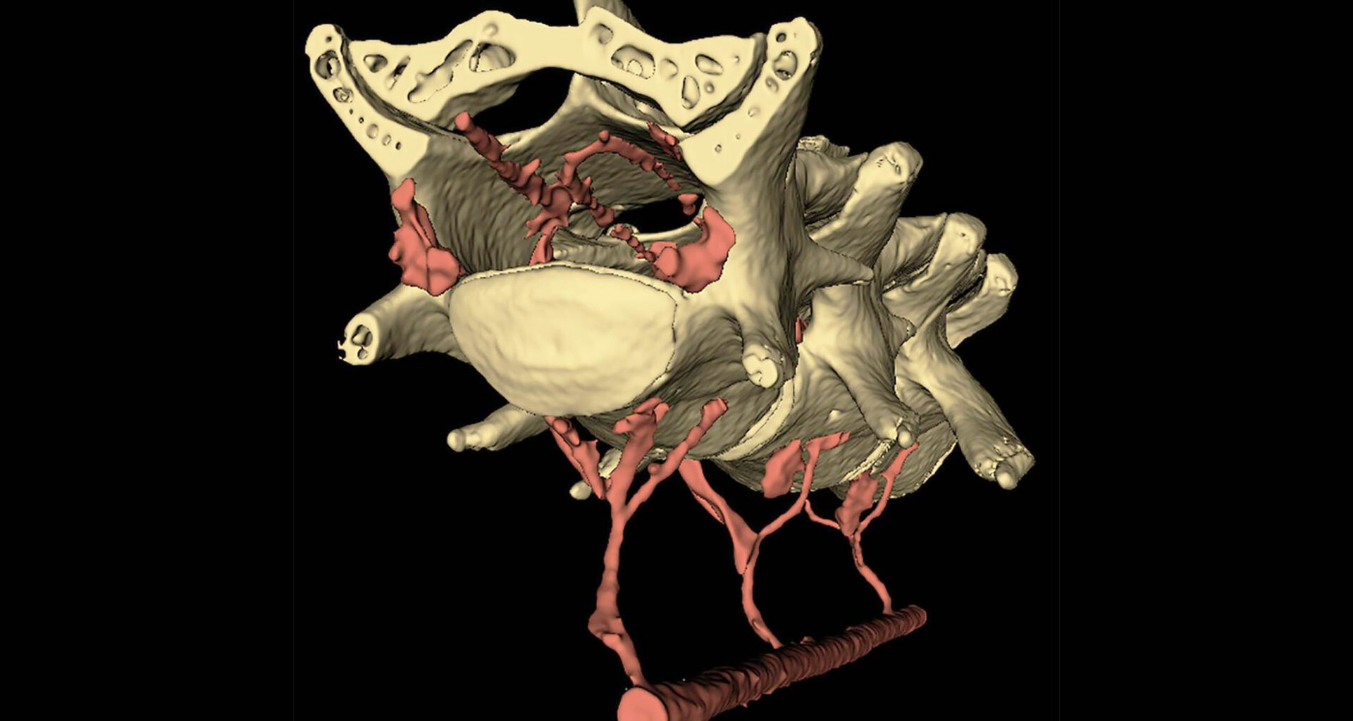
Using high-resolution 3D scans, the team mapped the blood vessels around each vertebra. They found valveless veins – which allow blood to flow in either direction – running through the lower vertebrae, linked through small openings to vessels in the abdomen.
That network had been described in humans but was never confirmed in mice. When the abdomen squeezes, blood is likely pushed through those openings into the spinal canal.
The added pressure appears to compress the fluid-filled sleeve around the spinal cord, sending a wave of cerebrospinal fluid – the clear liquid that cushions the brain and spine – forward into the skull.
A pressure cuff test
The researchers built a small, inflatable belt for lightly anesthetized mice. No walking, no voluntary contractions – just a gentle external squeeze on the abdomen, less than the squeeze of a blood pressure cuff.
Abdominal pressure alone made the brain move – forward, slightly sideways, the same direction as during walking. When the belt deflated, the brain returned to its resting position almost immediately.
“This suggests that abdominal pressure can rapidly and significantly alter the position of the brain within the skull,” Drew said.
Modeling fluid flow
Imaging fluid moving through brain tissue in real time isn’t yet possible. So the team built a computer model.
Francesco Costanzo, professor of engineering science and mechanics at Penn State, led the simulation work.
The result: the squeeze on the spinal cord drove fluid out of the brain and into the surrounding space several times faster than the brain normally produces its own fluid. A modest core contraction may routinely flush waste outward.
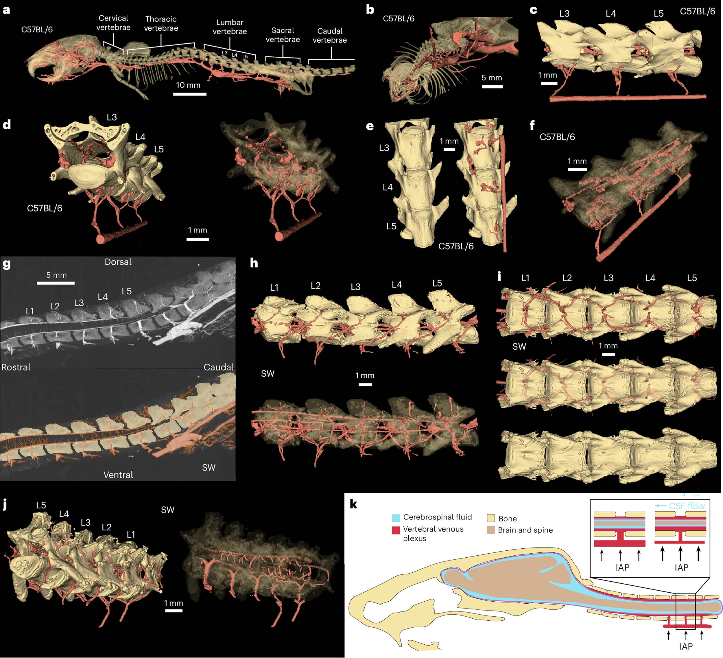 Using microCT scanning, which allows for high-resolution imaging of an organism’s internal structures, and other imaging techniques, researchers found that a network of veins serve as a mechanical connection between the abdominal cavity and the brain. Here, the veins in red run through the interior of the vertebrae and around the spine. Credit: Provided by Patrick Drew and team/Penn State. All Rights Reserved. Click image to enlarge.Opposite of sleep
That direction was the unexpected part. The brain’s glymphatic system – the waste-clearing pathway activated during sleep – pulls fluid into the brain along the outside of blood vessels while a person sleeps.
Until this study, no one had identified a mechanical reason why the awake brain might be doing the opposite. Those simulations show fluid flowing outward during waking hours – the reverse of what an earlier study on sleep and brain waste clearance described.
That helps explain a long-standing puzzle: tracers injected into the spinal fluid of awake mice don’t enter the cortex, while the same tracers travel freely during sleep. The two states seem to alternate – sleep for intake, waking for outflow.
Mouse brains are not human brains
All experiments were conducted in head-fixed mice, not humans, and the fluid flow findings come from simulation rather than direct imaging. Whether the same mechanism operates in people – and at what scale – remains to be tested.
What comes next
The study overturns a basic assumption: the brain is not mechanically isolated from the body. Abdominal contractions nudge it forward within the skull and may sweep waste fluid outward as they do.
That opens a new line of inquiry. Conditions that chronically alter abdominal pressure – such as obesity, extended bed rest, gastrointestinal disease – could quietly disrupt fluid clearance over time. Daily movement may help preserve it, consistent with research that links physical activity to better brain health.
“Our research shows that a little bit of motion is good, and it could be another reason why exercise is good for our brain health,” Drew said.
The study is published in Nature Neuroscience.
—–
Like what you read? Subscribe to our newsletter for engaging articles, exclusive content, and the latest updates.
Check us out on EarthSnap , a free app brought to you by Eric Ralls and Earth.com.
—–