

A 32-year-old Hispanic woman presented for evaluation of longstanding decreased vision in both eyes since birth. Prior to relocating to the United States, she had undergone a single eye exam at age nine by an ophthalmologist in Nicaragua, at which time she was told there was no treatment available to restore her vision. Her medical history was significant for migraines and moderate-to-severe sensorineural hearing loss. Notably, the patient had a fraternal twin sister with tetralogy of Fallot, hearing loss, decreased vision and developmental delays.
On examination, best-corrected visual acuity was 20/200 OD and 20/60 OS. Intraocular pressures were 13mm Hg OD and 12mm Hg OS. Pupils were round, equal and reactive to light with no relative afferent pupillary defect. Extraocular movements were full bilaterally without subjective diplopia or pain. Color vision testing with Ishihara plates was normal (11/11) OD and OS. Confrontation visual fields were full to finger counting bilaterally. Anterior segment examination was unremarkable.
Take the Retina Quiz1. Which of the following best describes the fundus features depicted in Figure 1?
a. Bone-spicule pigmentation.
b. Bull’s-eye maculopathy.
c. Cherry red spot.
d. Salt-and-pepper retinopathy.
2. Which structure is primarily affected to produce the characteristic fundus features seen in Figure 1?a. Choriocapillaris.
b. Nerve fiber layer.
c. Photoreceptors.
d. Retinal pigment epithelium (RPE).
3. What is the most likely diagnosis?
a. Congenital rubella syndrome (CRS).
b. Congenital stationary night blindness.
c. Congenital syphilis.
d. Congenital toxoplasmosis.
4. Which of the following is the classic triad of CRS?
a. Chorioretinitis, hydrocephalus, intracranial calcifications.
b. Hutchinson’s teeth, sensorineural hearing loss, interstitial keratitis.
c. Cataracts, congenital heart defects, sensorineural hearing loss.
d. Periventricular calcifications, microcephaly, sensorineural hearing loss.
5. The highest risk of developing ocular manifestations from this disease occurs during which trimester?
a. First trimester.
b. Second trimester.
c. Third trimester.
d. Both B and C.
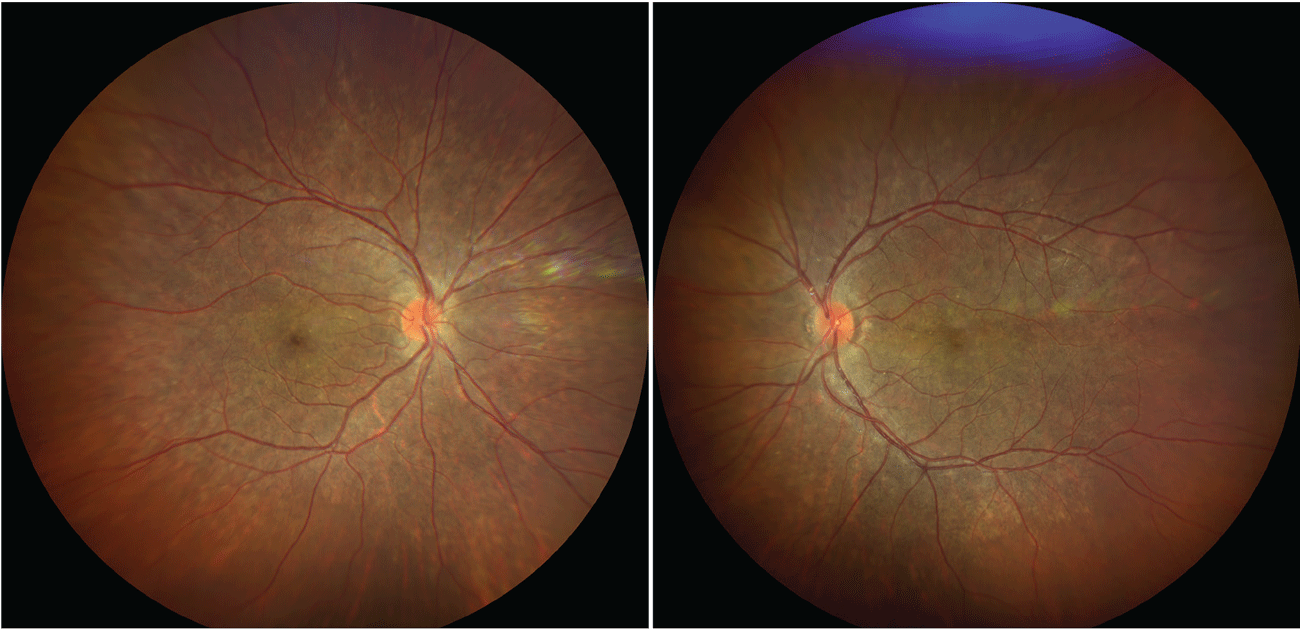
Fig. 1. Clarus fundus photographs OD and OS. Click image to enlarge.
Diagnosis
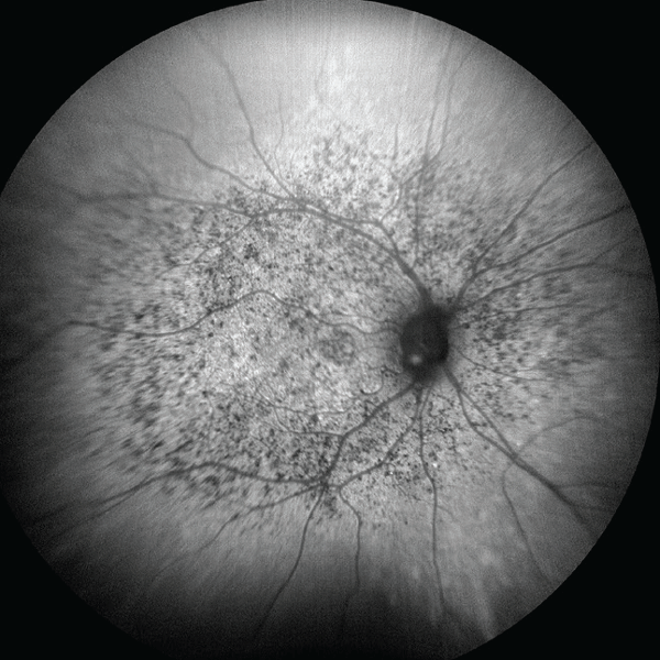
Fundus examination revealed a diffusely hypopigmented macula with scattered hyperpigmentary changes throughout the posterior pole and midperipheral fundus OU; notably, the retinal vasculature and optic nerve were unremarkable (Figure 1). Fundus autofluorescence demonstrated diffuse macular hypoautofluorescence corresponding with pigmentary changes seen clinically (Figure 2). Optical coherence tomography (OCT) demonstrated diffuse attenuation and discontinuity within the RPE (Figure 3). This clinical presentation was characteristic of salt-and-pepper retinopathy; a typical ocular manifestation associated with CRS.
DiscussionRubella, also known as German measles, is a viral infection that is transmitted primarily through respiratory droplets. It commonly presents with lymphadenopathy, arthralgia, fever and a maculopapular rash, though many patients are completely asymptomatic.1,2 Despite being highly contagious, rubella infections are typically mild and self-limited in children and adults.1 When infection occurs during pregnancy, however, consequences are much more devastating; transplacental viral transmission can result in fetal miscarriage, death or anomalies consistent with CRS. Risk to fetal development is greatest during the first 12 weeks of pregnancy (i.e., first trimester), when organ development is most critical.1,2History
Rubella was first described in the early 18th century by German physicians as a disease distinct from measles and scarlet fever, thereby becoming known as “German measles.”2 In 1941, Dr. Norman Gregg, an Australian ophthalmologist, published his landmark observation of congenital cataracts present in infants with CRS following maternal infection.1 In 1962, the rubella virus was isolated for the first time, and in 1964 through 1965 rubella swept across the United States in the last major epidemic outbreak. An estimated 12.5 million cases of rubella infection resulted in about 20,000 livebirths with CRS and thousands of additional miscarriages and neonatal deaths during the epidemic, sparking widespread efforts in vaccine development and public health immunization programs.1
The first dedicated rubella vaccine became available in the US in 1969, followed by the combined measles, mumps and rubella (MMR) vaccine in 1971.2 The MMR is administered at 12 to 15 months and again at four to six years of age, providing lifelong immunity.2 Widespread adoption of the vaccine led to a dramatic decline in rubella cases, and it was officially declared to be eradicated in the US in 2004, with fewer than 10 cases of rubella reported annually thereafter.2,3 While eradicated within the US, rubella remains common on the global stage, with an estimated 100,000 infants born with CRS each year.2 Despite the World Health Organization’s efforts to expand immunization access, rubella remains the leading vaccine-preventable cause of birth defects worldwide.
Pathophysiology
While the pathophysiology of CRS is not completely understood, it is thought to be multifactorial. The rubella virus is believed to cause noninflammatory necrosis, direct cytolysis and disruption of cellular mitosis within endothelial cells and cells of the chorionic epithelium.3,4 Viral interference of actin assembly also plays a role through inhibiting cell mitosis.3,4 By inhibiting cell division, the virus reduces the pool of precursor cells available for differentiation, thereby impairing organ development during critical periods of fetal growth. Upregulation of interferon and pro-inflammatory cytokines may further contribute to the clinical manifestations of CRS.3 Rubella infection during the first eight weeks of pregnancy carries a 50% fetal infection rate and the rate of in utero transmission falls below 10% by the 16th week.5
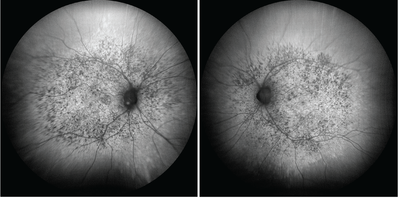
Fig. 2. Clarus fundus autofluorescence of OD and OS. Click image to enlarge.
Diagnosis in Newborns
In newborns, CRS can be confirmed by detection of rubella-specific IgM antibodies or by the persistence of rubella-specific IgG antibodies beyond six to 12 months of age (i.e., beyond the expected window of passive maternal antibody transfer.)11 Infants born with CRS may remain contagious for up to one year and are recommended to be screened for active viral shedding monthly after three months of age until two negative results are documented.2 Diagnostic accuracy declines as antibody levels wane, making serologic confirmation in children older than one year of age less straightforward. In adults with suspected CRS, diagnosis is typically presumed through careful history taking and the constellation of classic clinical findings.
Differential Diagnosis
The most important differential diagnosis of salt-and-pepper retinopathy to consider is congenital syphilis, which similarly presents with a range of systemic findings including Hutchinson’s teeth, saber shins, hydrocephalus, sensorineural hearing loss and saddle nose deformity.6 Unlike rubella-associated retinopathy, however, congenital syphilis is more commonly associated with signs of ocular inflammation—most notably interstitial keratitis.6
Additionally, rubella-associated retinopathy tends to be nonprogressive and symmetric, whereas congenital syphilis is more variable and progressive.6, 7 The differential diagnosis of pigmentary retinopathy should also include inherited retinal dystrophies and drug toxicity from phenothiazines (thioridazine) and antimalarials (hydroxychloroquine, chloroquine).
Ocular Manifestations
The classic triad associated with congenital rubella syndrome includes sensorineural hearing loss, congenital heart defects and cataracts.1,5 While congenital cataracts are among the most well-known ocular manifestation of CRS and characterized by dense opacification of the lens, pigmentary retinopathy is the most common ophthalmic feature of CRS, present in >50% of patients and typically bilateral.5 Congenital cataracts—present in about one-fourth of patients—occur as the crystalline lens is particularly vulnerable to viral injury during the first, when its epithelial cells are actively proliferating and differentiating into lens fibers.1
Pigmentary retinopathy is another common ocular manifestation of CRS and presents as multifocal RPE hyper/hypopigmentation predominantly throughout the posterior pole but extending into midperiphery.1,5 These features are generally stable and non-progressive throughout life. Rarely, however, RPE alterations may give way for macular neovascularization (MNV), requiring monitoring for this sequela.1,2,5,8 Additionally, progressive enlargement of RPE atrophy over time has been documented.7
Other, less common ophthalmic features of CRS may include microphthalmia (10% to 20% of patients with CRS), strabismus, nystagmus, congenital glaucoma and iris atrophy.1 Interestingly, histopathological analysis of patients with congenital glaucoma has demonstrated maldevelopment of the iridocorneal angle, and iris atrophy and hypoplasia are thought to result from poor development of the iris dilator muscle.1 Lastly, rubella has been implicated in the pathogenesis of Fuchs’ uveitis syndrome, with studies showing persistent rubella virus antibodies and genomic material present in the aqueous humor of affected eyes.9,10
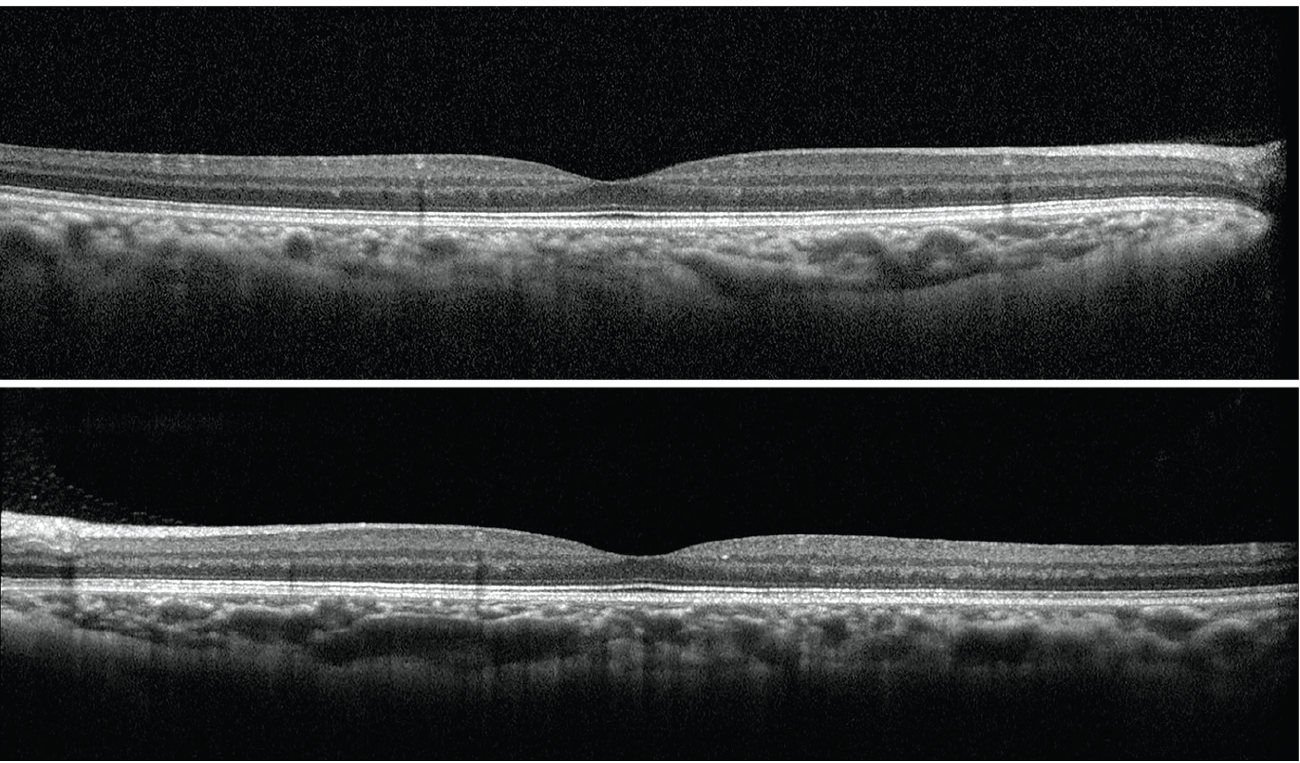
Fig. 3. Heidelberg OCT scans OD (top) and OS (bottom). Click image to enlarge.
Management and Prognosis
Salt-and-pepper retinopathy is generally stable and nonprogressive; however, annual dilated fundus exams are needed to monitor for development of MNV and enlargement of RPE atrophy.1,6 Advanced congenital cataracts may require surgical intervention and can be complicating when accompanied by microphthalmia or anterior segment dysgenesis.1 The rubella virus has also been shown to be present in the lens for prolonged periods after birth and thus may pose a theoretical risk of operating room personnel who are immunocompromised, unvaccinated or pregnant.1 Overall, management of CRS typically requires a multidisciplinary approach that also includes neurology, cardiology and audiology.1
Back to Our Patient
Our patient was referred to the retina service for monitoring and cardiology service for baseline comprehensive cardiac work up, as this had never been completed. Unfortunately, she was lost to follow-up and was a no-show to all her appointments.
Takeaways
While the ocular manifestations of congenital rubella syndrome are rarely encountered in contemporary practice, the last major epidemic included those of the Baby Boomer generation and thus must remain on the differential when examining patients with longstanding poor vision and pigmentary retinopathy. Furthermore, rubella remains a concern in other parts of the world and should also be considered in patients born abroad. It is important to remember the extraophthalmic manifestations of CRS including cardiac defects and sensorineural hearing loss and refer appropriately to other subspecialties.
Retina Quiz Answers
1: d, 2: d, 3: a, 4: c, 5: a
Dr. Aboumourad currently practices at Bascom Palmer Eye Institute in Miami. He has no financial disclosures.
Dr. Edwards is a recent graduate who completed an ocular disease residency at Bascom Palmer Eye Institute earlier this year.