
Optical coherence tomography (OCT) has transformed the diagnosis and management of retinal, optic nerve and anterior segment diseases. It is incorporated in evidence-based practice guidelines and pivotal clinical trials as the reference imaging modality for diagnosis, treatment decisions and monitoring. Spectral-domain OCT (SD-OCT) instruments have remained the mainstay office-based platform taking over the previous time-domain OCT (TD-OCT) option. Advancements in OCT technology have expanded its clinical applications, enabling imaging from the cornea to the peripheral retina.
Clinicians have several commercially available spectral-domain (SD) OCT models to choose from. For this article, four prominent ones will be described: Cirrus HD SD-OCT 5000 (Carl Zeiss Meditec), 3D OCT-1 Maestro2 (Topcon Healthcare), Spectralis SD-OCT (Heidelberg Engineering) and Optovue iVue80 (Visionix). This article outlines the basics of OCT imaging, the newest model updates, a guided data analysis of the different instruments’ metrics and how to interpret various measurements using each model.
Enhanced Depth Imaging (EDI-OCT)
This technology positions deeper structures near the zero-delay line. This technique is now built into many SD-OCT instruments and provides improved visualization of the choroid, lamina cribrosa and scleral structures.
Visualization of the choroid-scleral junction enables quantitative choroidal thickness measurements. Increased choroidal thickness, characterized by a dilated Haller’s layer with a thinned inner Sattler’s layer and choriocapillaris, is a key diagnostic feature of pachychoroid spectrum disorders.1 Conversely, a thinner choroid can assist in differentiating age-related macular degeneration from macular dystrophies such as adult-onset foveomacular vitelliform dystrophy.2 Reduced choroidal thickness is also associated with pathologic myopia and correlates with axial elongation, severity of myopic maculopathy and an elevated risk of progression to atrophic and neovascular complications.3,4
Peripapillary choroid thickness has emerged as a potential biomarker in both optic nerve and retinal diseases. Peripapillary choroid thickness is increased in pachychoroid spectrum disorders, such as peripapillary pachychoroid syndrome, and decreased in optic neuropathies such as glaucoma, particularly normal-tension glaucoma.5,6 EDI-OCT, with its deeper signal penetration, improves visualization of deep optic nerve head structures and enables in vivo assessment of the lamina cribrosa (LC). Lamina cribrosa parameters that have been studied using EDI-OCT include LC thickness, anterior LC depth, LC curvature, focal LC defects and prelaminar neural tissue thickness. Although these LC parameters provide quantitative measures of glaucomatous deformation, cupping and remodeling, their utility remains primarily research and as adjunctive clinical tools. Currently, no commercially available FDA-approved OCT software offers automated LC analysis.7
The Optic Disc Drusen Studies Consortium recommends EDI-OCT as its preferred diagnostic modality for the detection of optic disc drusen (ODD). On EDI-OCT, ODD is located anterior to the LC inside Bruch’s membrane opening and appears with a hyporeflective core and full or partial hyperreflective margin.8 In a comparative study of 205 eyes, EDI-OCT demonstrated the highest sensitivity and accuracy relative to fundus autofluorescence (FAF), B-scan ultrasonography and fundus photography.9 Optical coherence tomography can also differentiate ODD from peripapillary hyperreflective ovoid mass-like structures (PHOMS), which are nonspecific markers of axoplasmic stasis of dislocated nerve fiber layer axons. On OCT, they appear as hyperreflective structures located above the Bruch’s membrane.10
EDI-OCT also assists in differentiating pseudopapilledema from true papilledema. OCT biomarkers that distinguish papilledema include peripapillary Bruch’s membrane and retinal pigment epithelium (RPE) configuration, which may exhibit anterior displacement or inward angulation towards the vitreous in response to elevated intracranial pressure. There is also the presence of peripapillary wrinkles, retinal (Paton’s) folds and choroidal folds.11,12 Additional OCT biomarkers, such as increased peripapillary retinal nerve fiber layer (RNFL) thickness and optic nerve head volume, are typically observed in papilledema.13
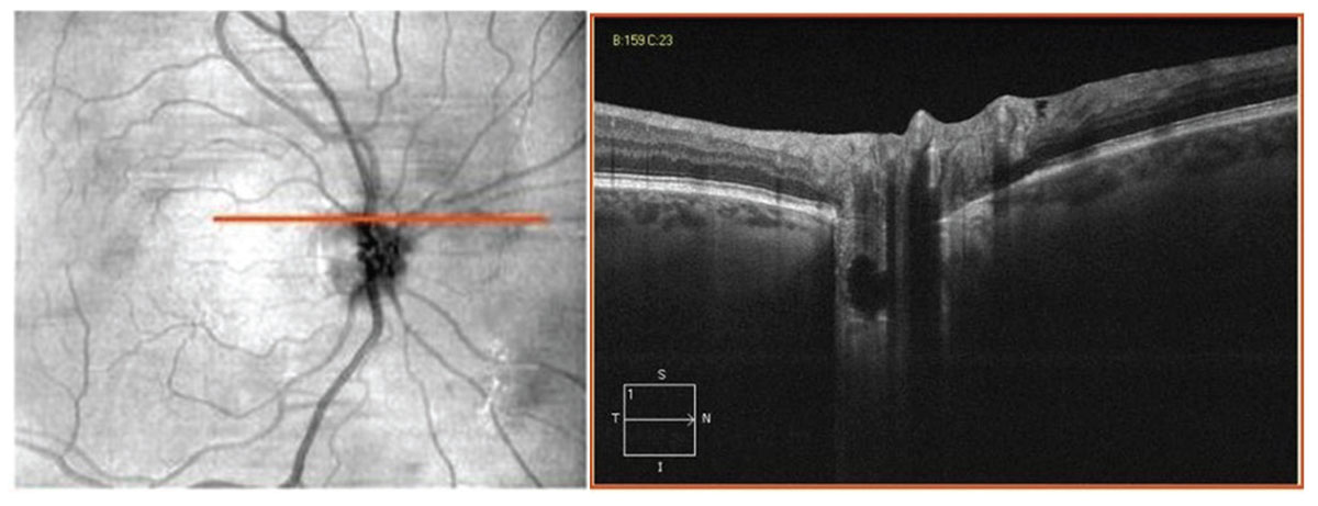
Optic disc drusen imaged with EDI-OCT. Click image to enlarge.
OCT Angiography (OCT-A)
This noninvasive, dye-free, depth-resolved imaging modality uses motion contrast generated by erythrocyte movement between repeated B-scans acquired at the same location. It is primarily used in clinical practice for visualization and assessment of the microvasculature within the retinal capillary plexuses, choroid and choriocapillaris.
In patients with diabetes, OCT-A imaging enables the detection of early subclinical microangiopathy, as demonstrated by microvascular alterations such as foveal avascular zone enlargement and reduced vessel density, even before retinopathy becomes visible on ophthalmoscopy. This technology can also identify retinal and optic disc neovascularization, facilitating the detection and monitoring of progression or regression in response to treatment. Additionally, OCT-A assists in differentiating intraretinal microvascular abnormalities from retinal neovascularization and provides quantitative assessment of retinal ischemia in diabetic retinopathy, which correlates with disease severity and helps predict progression.14
As a diagnostic tool, OCT-A has emerged as an important imaging modality for detecting and characterizing macular neovascularization (MNV) in age related macular degeneration (AMD). Unlike fluorescein angiography (FA), OCT-A allows visualization of the retinal and choroidal vasculature without obscuration from dye leakage. The combined use of en face OCT-A and cross-sectional OCT-A enhances the sensitivity for MNV detection.15
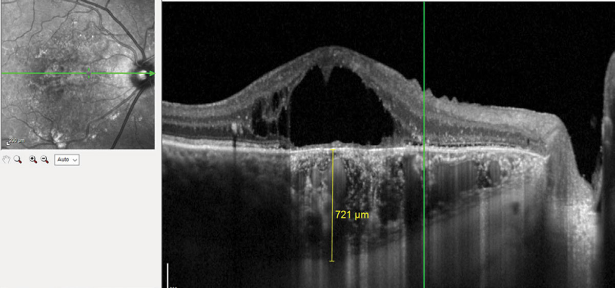
Chronic central serous chorioretinopathy with pachychoroid imaged with EDI-OCT. Click image to enlarge.
This modality can also help classify the three subtypes of MNV (type 1, 2 and 3) that can predict both response and complication risk. Type 1 MNV typically requires sustained anti-VEGF dosing. It has been found that only 11.9% of the intravitreal anti-VEGF concentration reaches the sub-RPE compartment, therefore a higher number of injections is required to treat type 1.16 In type 2 MNV, the lesion is in the subretinal space and is associated with worse baseline visual acuity but demonstrates a rapid response to anti-VEGF therapy. However, type 2 MNV is more prone to developing subretinal fibrosis.17 Type 3 MNV, also called retinal angiomatous proliferation, generally responds to anti-VEGF therapy but has the highest risk for development of outer retinal atrophy.18
For identifying and monitoring nonexudative MNV, OCT-A demonstrates high sensitivity. This type of MNV is a treatment-naive neovascular network without evidence of signs of subretinal or intraretinal fluid. On structural OCT, it presents as a shallow irregular RPE elevation with nonhomogeneous internal reflectivity. The shallow irregular RPE (SIRE) sign is defined by an RPE elevation measuring ≥1,000µm in length and <100µm in height and has been associated with a higher predictive value for nonexudative MNV. OCT-A is the preferred modality for detecting nonexudative MNV, demonstrating a sensitivity of 81.8% and a specificity of 100%. This identification is crucial due to this type of MNV’s risk for conversion to exudative AMD. The two-year risk of conversion to exudation has been reported to be up to 13.6-fold higher in these eyes.19
Assessment of optic neuropathies also benefits from the detailed imaging capabilities of OCT-A, particularly for the radial peripapillary capillary plexus and superficial vascular complex. In glaucoma, OCT-A detects loss of these structures and has been shown to correlate with both glaucoma severity and structural RNFL thinning. Comparative analyses of the diagnostic efficacy of OCT-A and structural OCT in head-to-head studies have produced inconclusive findings. The American Academy of Ophthalmology (AAOph) states that OCT-A may complement visual field and structural OCT measurements, but additional studies are needed to evaluate the device’s parameters on predicting glaucoma onset and progression.20
Limitations of OCT-A include the inability to detect leakage, flow-detection threshold causing poor visualization of slow-flowing lesions, segmentation errors resulting from pathology and narrow normative database with quantitative metrics, such as vessel density, perfusion density and foveal avascular zone measurements.
Emerging applications of OCT-A include anterior-segment and widefield imaging. With swept-source technology (SS-OCT), dedicated anterior-segment OCT-A can penetrate deeper to image cornea, iris and angle vasculature. Swept-source OCT’s faster scan rates can also reduce motion artifacts and image mid-peripheral and peripheral retina to detect peripheral non-perfusion and neovascularization.
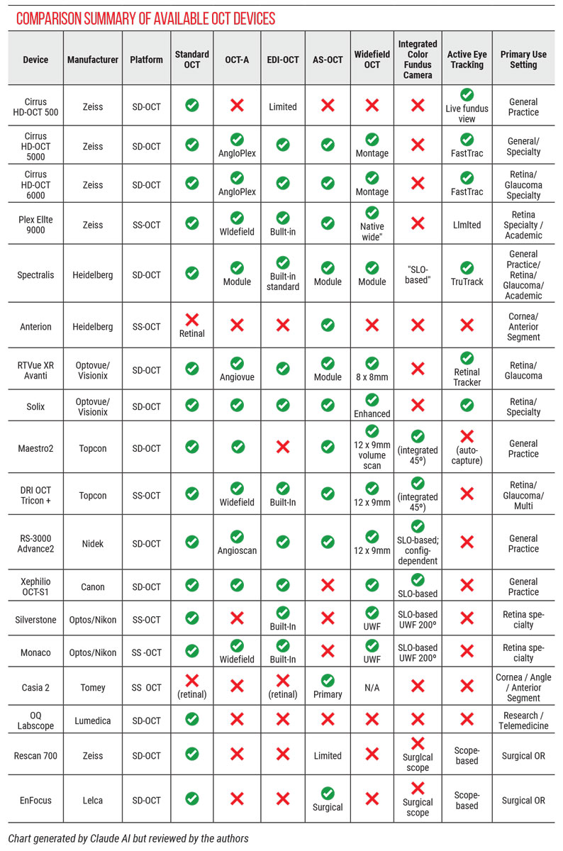
Click table to enlarge.
Swept-source OCT
This technology uses a rapidly tunable laser light source, enabling faster scan rates and longer wavelengths relative to SD-OCT. With scan speeds of up to 400,000 A-scans per second, SS-OCT facilitates denser and more complex scan patterns, as well as widefield image acquisition with reduced motion artifacts compared to SD-OCT. The longer wavelength used by SS-OCT, approximately 1,050nm for retinal imaging compared to 840nm for SD-OCT, permits deeper tissue penetration and allows for detailed choroidal imaging without the need for EDI-OCT. For anterior-segment imaging, SS-OCT operates at even longer wavelengths, approximately 1,300nm.
Similar to EDI-OCT, SS-OCT enhances the visualization of choroidal and optic nerve head structures, aiding in the diagnosis and management of conditions such as pachychoroid spectrum disorders and ODD. However, the SS-OCT’s faster scan speeds confer additional advantages, enabling the acquisition of denser and more complex scanning patterns and enhanced anatomical detail. SS-OCT can simultaneously visualize the vitreous, retina and choroid, a capability that distinguishes it from EDI-OCT, which typically sacrifices vitreous detail due to positioning near the zero-delay line.
Widefield (WF) OCT and ultra-widefield (UWF) OCT are also enhanced with SS-OCT compared to SD-OCT, primarily due to the faster A-scan acquisition speed of SS-OCT. Additionally, the 1,050nm wavelength used in SS-OCT minimizes light scattering and significantly reduces sensitivity roll-off with increasing imaging depth, a feature that is crucial for accurate imaging of the curved peripheral retina during both of these OCT scans.21
Faster acquisition rates in SS-OCT also improve OCT-A scans due to fewer motion artifacts and the ability to scan a larger area. These improvements are particularly relevant in OCT-A, which relies on motion contrast from repeated B-scans at the same location. Larger fields, such as 12 × 12mm or 15 × 9mm, allow SS-OCT to extend assessment of blood flow and vessel structure to outside the vascular arcades. Deeper tissue penetration further improves visualization of the choriocapillaris and large choroidal vessels in Haller’s layer.
Despite these advantages, the clinical adoption of SS-OCT remains limited due to higher costs and a relatively small number of commercially available instruments, many of which are intended for research use or specifically designed for widefield OCT or anterior-segment OCT (AS-OCT) applications.
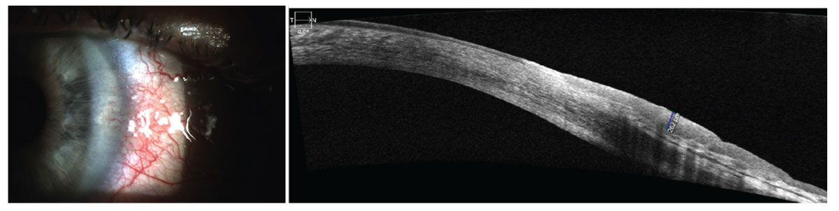
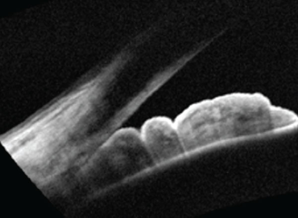
Ocular surface squamous cell neoplasia imaged with AS-OCT. There is a thickened and hyperreflective epithelial layer with an abrupt transition from normal to abnormal epithelium. Click image to enlarge.
Anterior Segment OCT
This is a valuable noninvasive imaging modality for the evaluation of the ocular surface, cornea, anterior chamber, angle and lens. Often described as an “optical biopsy,” AS-OCT enables detailed assessment of conjunctival and corneal lesions. This technology provides objective and reproducible qualitative and quantitative metrics and facilitates imaging through ocular opacities that may obscure visualization with conventional slit-lamp biomicroscopy. Early TD-OCT instruments were dedicated exclusively to either anterior segment or posterior segment imaging. In contrast, several SD-OCT platforms, originally optimized for retinal and optic nerve imaging, now incorporate high-powered lenses or lens attachments that enable AS-OCT imaging. Although these instruments use an 840nm wavelength optimized for retinal imaging, their expanded AS-OCT capabilities have enabled anterior segment imaging without the need for an additional OCT system. Incorporating both SD-OCT and SS-OCT technologies, modern AS-OCT devices offer enhanced depth penetration and support advanced imaging capabilities, including volumetric (3D) acquisition.
One of the primary clinical applications of AS-OCT is in glaucoma. This device can provide direct visualization and quantification of the anterior chamber angle for angle-closure diagnosis and risk stratification. It is especially advantageous in patients who are unable to tolerate gonioscopy, providing objective measurements such as angle-opening distance, trabecular-iris space area and angle recess area to assess angle-closure risk. The Zhongshan Angle Closure Prevention (ZAP) trial found that AS-OCT biometric parameters were predictive of progression from primary angle-closure suspect to primary angle closure.22
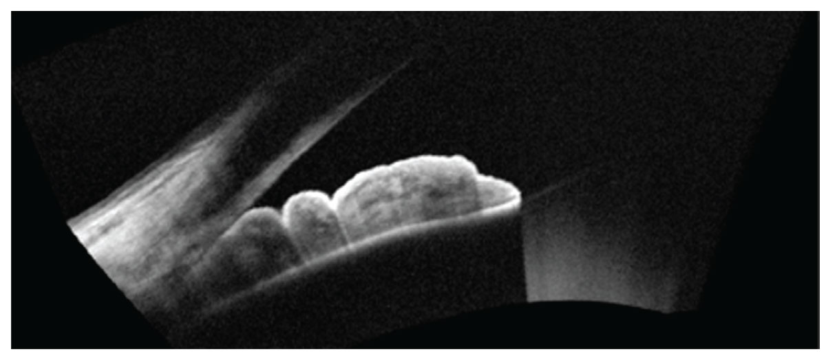
Iridocorneal contact imaged with AS-OCT. Click image to enlarge.
In addition, the use of infrared light in AS-OCT allows for imaging in complete darkness, thereby avoiding light-induced pupillary constriction. According to the AAOph Preferred Practice Patterns, AS-OCT serves as a valuable adjunct to gonioscopy and is particularly useful when gonioscopy cannot be performed.23
Other clinical applications of AS-OCT in glaucoma include the evaluation of filtering blebs following glaucoma surgery. It also enables the differentiation between functioning and non-functioning blebs, facilitating the early detection of filtration failure, often earlier than slit-lamp examination.24
Additionally, AS-OCT can image minimally invasive glaucoma surgery (MIGS) devices. A systemic review of this type of imaging in MIGS concluded that it can identify outflow pathways, document implant location, evaluate surgical endpoints and detect postoperative complications.25 AS-OCT also provides cross-sectional imaging of both the anterior and posterior corneal surfaces, generating pachymetry maps. It is a highly reproducible noncontact alternative to ultrasound pachymetry for central corneal thickness measurement.
Pachymetry maps acquired with AS-OCT are useful in refractive surgery screening, keratoconus detection and staging, evaluating corneal edema and endothelial disease and post-keratoplasty monitoring. Anterior-segment OCT epithelial thickness maps can be used to detect subclinical and forme fruste keratoconus. A characteristic doughnut pattern of localized epithelial thinning over the apex of the cone, surrounded by an annulus of epithelial thickening, can be seen on AS-OCT. This epithelial remodeling can mask early stromal changes, making them difficult to visualize on corneal topography.26
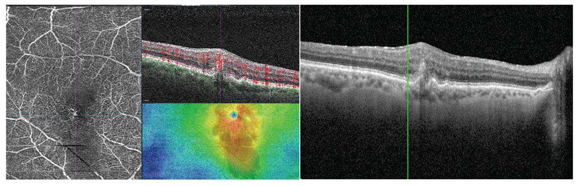
Type 3 MNV imaged with OCT-A with intraretinal neovascularization. Click image to enlarge.
Anterior segment OCT is a valuable tool in the assessment of ocular surface squamous neoplasia (OSSN). Anterior segment OCT aids in diagnosis by differentiating OSSN from clinically similar lesions and by distinguishing intraepithelial from invasive disease. Additionally, AS-OCT aids in guiding surgical margin determination and monitoring response to topical chemotherapy as well as detecting recurrence. On AS-OCT, OSSN presents as a thickened, hyperreflective epithelium with an abrupt transition between normal and pathological tissue. These imaging characteristics help distinguish OSSN from lesions such as pterygium, pseudopterygium and pinguecula, which appear significantly thinner on AS-OCT. Such findings are consistent with corresponding histopathology findings.27,28
Other clinical applications of AS-OCT include objective quantification of anterior chamber cells and flare in the diagnosis and management of anterior uveitis, as well as detailed imaging of the lens-to-eye relationship in the fitting of rigid gas-permeable (RGP), scleral and orthokeratology lenses. In scleral lens fitting, AS-OCT can be used to objectively measure the fluid reservoir for central clearance, limbal clearance and peripheral landing zone.29
Swept source AS-OCT instruments specifically designed for anterior-segment imaging use longer wavelengths (1,050nm to 1,310nm), which allow for improved visualization of the deep angle structures compared to SD-OCT devices. In addition to anterior segment imaging, these devices often integrate eye biometry and corneal topography capabilities. However, due to their high cost, dedicated swept-source AS-OCT systems are typically reserved for multi-subspecialty or high-volume anterior segment centers, particularly those specializing in cataract and refractive surgery. Limitations of AS-OCT include the inability to visualize structures posterior to the iris, lack of standardization and limited automated analysis.
WF and UWF OCT
These imaging modalities extend the field of view beyond the posterior pole, enabling visualization of the mid-peripheral and peripheral retina. Currently, most conventional OCT scans only image the posterior pole. However, some OCT instruments allow for image montaging, capturing multiple scans and merging them to extend outside the posterior pole. Other OCT instruments have WF add-on lenses that expand the scan field, allowing a single capture of the optic nerve, macula and beyond the vascular arcades.
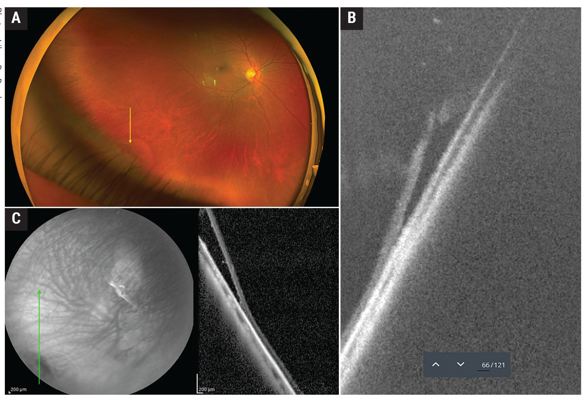
Shallow retinal detachment associated with retinal break imaged with UWF OCT (A and B)and WF OCT (C). Photos: Lilyan Do Bambao. Click image to enlarge.
Clinical applications of WF and UWF OCT imaging include detection of peripheral retinopathy (atrophy, neovascularization), peripheral vitreoretinal traction (breaks, detachment), peripheral retinal degeneration (retinoschisis) and peripheral lesions. By imaging peripheral retinal degenerations and characterizing the vitreoretinal interface, clinicians can stratify these lesions by their risk of developing rhegmatogenous retinal detachment. In a series of eyes with peripheral retinal degenerations including lattice degeneration, snail track lesions and peripheral retinoschisis imaged using UWF OCT, the authors found that OCT enhanced diagnostic capability. Specifically, UWF OCT enabled earlier identification of characteristic features such as vitreoretinal traction, full-thickness retinal breaks and subretinal fluid, which were not readily apparent on binocular indirect ophthalmoscopy.30
Widefield and UWF OCT can also help differentiate diagnostically ambiguous peripheral lesions. On UWF OCT, it can separate retinoschisis from retinal detachment from schisis-detachment, which can be difficult to distinguish on binocular indirect ophthalmoscopy.31
Emerging clinical applications of WF OCT-A and UWF OCT-A include imaging of peripheral retinal ischemia and detection of peripheral retinal neovascularization. This is particularly helpful in conditions like diabetic retinopathy, where the vision-threatening features can be outside the Early Treatment Diabetic Retinopathy Study (ETDRS) 7 fields and missed by conventional narrow-field OCT-A.32
Limitations of WF and UWF OCT include peripheral image quality degraded by optical aberrations, media interfaces, poor tracking and motion and montage artifacts.
Future Directions
Potential paths of OCT technology to explore include developing higher-speed instruments that enable UWF and functional extensions such as adaptative-optics OCT; miniaturization for handheld, home or surgery use; and pervasive artificial-intelligence-driven analysis.33,34
OCT is now a standard imaging modality in eye care. A comprehensive understanding of OCT technology and its expanding clinical applications is essential for delivering optimal eye care.
Dr. Vien is the director of optometry clinic operations and co-residency supervisor at the VA Palo Alto Health Care System. He is an associate clinical professor at Herbert Wertheim School of Optometry & Vision Science at UC Berkeley and State University of New York College of Optometry. He is also an assistant clinical professor at the Southern California College of Optometry at Marshall B. Ketchum University.
Dr. Yang is the chief of optometry and co-residency supervisor for the VA Palo Alto Health Care System. He is a clinical professor at Herbert Wertheim School of Optometry & Vision Science at UC Berkeley. He is also the co-chair of the VA National Optometry Contact Lens Workgroup.