

Introduction
The sport of rugby is played by roughly 9 million athletes around the world, attracting large amounts of spectators.1 Due to the nature of this collision sport, participants are frequently tackling or being tackled, leaving them exposed to repetitive head impacts (RHIs). Research concerning RHIs has been gaining popularity due to the potential for long-term neurological deficits in the absence of immediately noticeable symptoms which are typically experienced following sport-related concussion.2–4 With this, identification of subtle alterations and interventions to prevent or limit neurological dysfunction are being explored. One such approach is supplemental intervention, representing a simple, unique method of addressing this growing concern.
Rugby Background
Tackles are the primary source of exposure to RHIs, with tacklers themselves at a greater risk of repetitive head impact exposure and concussive injury compared to the player being tackled.5–9 Moreover, players at forward positions appear to sustain a greater number of RHIs compared with those playing in back positions.10 RHIs in rugby are substantial at each level, with 4718 and 5345 tackles observed across 20 matches for U18 and U22 players, respectively.8 Professional rugby players have been shown to sustain as many as 15 RHIs in one minute at their peak. When looking at a 10-minute time frame, an average of 5 RHIs were sustained, equating to 40 RHIs per game.11 While data concerning exposure to RHIs in rugby practice are lacking, practices are conducted multiple times per week – often with full contact – presenting greater risk for exposure to potentially harmful RHIs.
Visual and Cognitive Deficits Associated with RHIs
Visual dysfunction following RHIs can manifest in multiple forms. Structural changes to retinal tissue including the ganglion cell complex (GCC) and retinal nerve fiber layer (RNFL) result in impulse transmission deficiencies while also leaving the individual at an increased risk of further structural damage including macular holes and retinal detachments.12–14 Continued structural change will eventually lead to functional deficits in the form of decreased contrast sensitivity (CS) and visual acuity, particularly once 25–35% of the GCC is lost.15–17 Further, inconsistent and slower visual processing speed, known as critical flicker-fusion frequency (CFF), has been seen in individuals with head injury.18 Faster CFF along with higher levels of macular pigment optical density (MPOD) are correlated with higher cognitive scores.19 Thus, protecting the visual system in turn protects cognitive functioning. While these changes typically occur over longer periods of time, transient visual dysfunction has been represented by increased near point of convergence (NPC)20,21 and slower King-Devick testing times22 immediately following exposure to RHIs. Similarly in cases of diagnosed concussion among rugby players, oculomotor deficits are present immediately and 30 days post-injury even when determined to be clinically asymptomatic.23,24 These findings reflect subclinical neuronal damage which may otherwise go unnoticed until a threshold of degradation is reached, supporting the need for frequent evaluation and the identification of protective measures.
Blood Biomarkers Associated with RHIs
Blood samples are popular in sport-related head injury research as they possess potential to provide swift insight into injury status and associated severity. Glial fibrillary acidic protein (GFAP) has been shown to increase in-season following exposure to RHIs in football players,25 from pre- to post-season in football players,26 and in deceased former football players and boxers.27 Further, combat sports athletes from boxing and mixed martial arts have shown increasing GFAP levels over time.28 Ubiquitin C-terminal hydrolase-L1 (UCH-L1) has been shown to increase immediately following exposure to RHIs in asymptomatic football players, and at post-season assessment.29,30 Although there is robust evidence for UCH-L1 elevation following exposure to RHIs, chronic elevation is not clear.31 Neurofilament light chain (NF-L) is an indicator of axonal injury32,33 and has been found at higher levels in football, hockey, and rugby athletes compared with healthy controls.26,32–34 Tau is detectable in both the acute and chronic phases of exposure to RHIs and brain injury, with research findings among football, hockey, and boxing athletes, as well as military veterans.32,33 Support for the assessment of multiple blood biomarkers is promising. GFAP, UCH-L1, NF-L, and Tau were found to be increased compared to baseline levels following sport-related concussion from 0–28 days post-injury,34 and for GFAP, NF-L, and Tau following exposure to RHIs when compared to controls.33
Macular Pigment Carotenoids
Macular Pigment Carotenoids (MPCs) are xanthophylls stored in the brain and eyes of humans, particularly in the macula of the retina.35,36 Humans are unable to synthesize MPCs and are thus required to obtain them through diet, presenting issues with their bioavailability due to the presence of cellulose.36,37 MPCs consist of lutein and zeaxanthin which are found in green-leafy vegetables, fruits, and egg yolk, while meso-zeaxanthin is believed to be found in a variety of cold-water fish and known to be a lutein metabolite.35 A combination of all three MPCs are argued to be best for optimization of expected benefits.36,38 Their well-known functions consist of increased processing speed, glare and blue light reduction, increased MPOD, improved contrast sensitivity threshold, and slowing ocular disease progression such as macular degeneration and cataracts.35,39–41 MPOD is a measure of the concentration (i.e. density) of lutein, zeaxanthin, and meso-zeaxanthin, defined by the amount of short-wavelength, blue light that is absorbed by a given concentration of each MPC in the macula. Blue light causes photochemical injury to the retina via cell and photoreceptor apoptosis,42–44 oxidative stress, and DNA damage.45 Thus, higher levels of MPOD are useful in protecting the tissue from associated blue light damage while also reducing the risk of age-related macular degeneration.46 Additional functions gaining interest in research protocols include their ability to protect brain and retinal tissues from oxidative stress and inflammation.35,37,47–49 Preclinical studies utilizing MPCs following head injury have revealed their ability to limit inflammation and oxidative stress while maintaining motor and cognitive functioning in rodent models.50–54 However, clinical studies utilizing human subjects have not been conducted and must be undertaken to limit this gap in knowledge concerning effectiveness of MPCs as limiting agents for outcomes associated with exposure to RHIs.
Omega-3 Fatty Acids
Omega-3 fatty acids eicosapentaenoic acid (EPA) and docosahexaenoic acid (DHA) are essential structural components of cellular membranes55 but are gaining popularity for their potential to treat health conditions influenced by inflammation and oxidative stress.56 Synthesis of EPA and DHA is inefficient in humans, meaning they must be obtained from the diet or supplements. Common sources of EPA and DHA include fish oils from salmon, tuna, and mackerel.56,57 While there is uncertainty surrounding daily intake requirements, it is evident that athletes are well below the daily recommendation and dietary intake alone makes it difficult to meet daily recommendations, signifying the need for a supplemental approach.58 EPA and DHA are stored in the brain and retinal tissue, with EPA quickly metabolized resulting in greater storage of DHA in neural tissue.59 Consuming greater quantities of EPA and DHA is associated with lower rates of chronic diseases characterized by inflammation, with DHA being shown limit axonal damage following brain injury.58,59 To combat inflammation, EPA and DHA accumulate in membranes of cells associated with inflammation.59 Upon activation of these cells, EPA and DHA are released and involved in the synthesis of lipid mediators utilized in the resolution of inflammation through the reduction of cellular activation, ultimately reducing the production of cytokines and enzymes accompanying inflammatory processes.56 Supplementation exhibits increased serum antioxidant capacity through an increase in antioxidant enzymes which assist in the reduction of free radicals and scavenging of reactive oxygen species (ROS).60,61
Synergy Between Macular Pigment Carotenoids and Omega-3 Fatty Acids
Bioavailability concerning nutrients of interest are discussed far less than they should be, often resulting in a misunderstanding of how to best optimize nutrition. Consumption of omega-3 fatty acids with carotenoids has been argued to result in greater bioavailability.62 As both MPCs and EPA + DHA selectively accumulate in brain and retinal tissue, a transport link is plausible. One study found DHA supplementation to increase lutein uptake into the macula, supporting such a link.63 In terms of their similar antioxidative capabilities, supplementation with both MPCs and EPA + DHA is believed to augment antioxidant effectiveness, but more research is needed before substantial claims can be made.60 Although omega-3 supplementation has been associated with decreased platelet aggregation, it’s prior association with oxidative damage due to extended bleeding times has been contested.64 Additionally, it represents well-documented anti-inflammatory utilities.65 Thus, omega-3 supplementation should be combined with additional antioxidants, such as MPCs to increase the likelihood of favorable outcomes.66 Studies evaluating supplementation with both MPCs and omega-3 fatty acids are limited but indicate significant cognitive benefits. A beverage with 125 mg DHA and 6 mg lutein improved working memory, reaction time, processing efficiency, verbal fluency, rate of learning, and learning efficiency in active-duty airmen vs placebo.67 Another study evaluating a combination of 12 mg lutein, 0.5 mg zeaxanthin, and 800 mg DHA found improved verbal fluency and memory, rate of learning, and learning efficiency in a population of older women vs those supplementing with either MPCs or omega-3 fatty acids on their own.63 As there is an apparent synergistic effect between MPCs and EPA + DHA benefitting shared accumulation sites, ocular health, cognition, and physiological processes, research employing otherwise healthy populations exposed to sustained RHIs is reasonable.
Neurological dysfunction following exposure to RHIs is a major public concern requiring a solution. MPCs and omega-3 fatty acids provide a wide range of benefits to the visual system, cognition, as well as limiting inflammatory and oxidative insult in healthy and diseased populations. However, their use in human subjects following exposure to RHIs has not been considered. Therefore, the purpose of this study was to a) assess both structural and functional integrity of the visual system of athletes at high risk of exposure to RHIs over a competitive season, and b) determine the effectiveness of a supplemental intervention of MPCs with EPA + DHA versus placebo in attenuating structural and functional visual changes following exposure to RHIs among collegiate rugby players from pre- to post-season. As a secondary aim we explored whether supplementation with MPCs and EPA + DHA influenced common blood biomarkers associated with neurodegeneration.
Materials and Methods
Design and Setting
This double-blind randomized placebo-controlled study consisted of male and female collegiate rugby players at Penn State University. All athletes provided written informed consent prior to participation following an explanation of the nature and possible consequences of the study. This study was approved by the Institutional Review Board at The Pennsylvania State University and complies with the Declaration of Helsinki. This study is registered as clinical trial NCT06270680.
Population
Participants were recruited during 2024 pre-season team meetings. All participants were at least 18 years of age and competing for men’s or women’s rugby 15’s teams at Penn State University. No participants reported concussion diagnosis within the last calendar year or diagnosis of an ocular or neurological disease.
Supplementation
The supplement and non-differentiable placebo were provided by MacuHealth LLC (Bloomfield Hills, MI, USA) as softgel capsules. The supplement contained 10 mg lutein, 2 mg zeaxanthin, 10 mg meso-zeaxanthin, 250 mg DHA, and 50 mg EPA (commercially available as Vision Edge Pro). The placebo contained 380 mg of sunflower oil. Packaging of bottles provided to participants were identical for supplement/placebo. Randomization of supplement/placebo bottles and the associated subject number occurred at MacuHealth LLC using =SORTBY(SEQUENCE(50),RANDARRAY(50)) excel code to return a random, non-repeated list of numbers in a column. Supplement or placebo designation was decided upon using returned even (placebo) and odd (supplement) numbers which were ordered top to bottom from subject 1 onwards. Participants were assigned a bottle with their corresponding subject number in the order in which their appointments were scheduled at pre-season testing. The institutional research team were masked to the supplement vs placebo condition, as only MacuHealth LLC, who conducted the randomization knew the identity of the supplement and placebo throughout the duration of the study. MacuHealth LLC maintained zero contact with participants. Participants were instructed to take one pill per day, ideally with a meal. Participants were informed that they could take two pills in one day if they had missed the previous dose. Compliance was monitored by instructing participants to complete a log sheet consisting of date and time following each dose.
Procedures
Participants completed pre- and post-season testing consisting of OCT scans, CS testing, CFF, MPOD, skin carotenoid concentration measurements, and a venous blood draw in a laboratory setting. Demographic and sport-specific data were collected and are presented in Table 1. Following pre-season testing, participants were provided with their supplement package and a log sheet to record intake. Researchers were unblinded following statistical analysis.
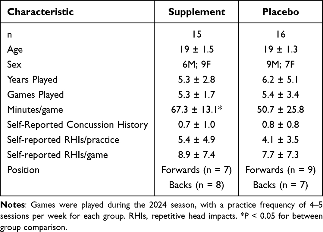
Table 1 Subject Demographic Information
Optical Coherence Tomography
The Optovue iScan 80 spectral-domain optical coherence tomography (OCT) machine was used to procure high-resolution images of retinal tissue consisting of GCC (superior/inferior quadrants), RNFL (superior/inferior quadrants), and RM (superior/inferior/nasal/temporal/central quadrants) average thickness in both the left (OS), and right (OD) eyes. This device uses the internal limiting membrane as the inner retinal border and the interdigitation zone as the outer retinal border. The same operator performed all scans to prevent operator variability.
Contrast Sensitivity Testing
The CTS contrast sensitivity vision testing system by M&S Technologies required subjects to complete 8 trials of best-corrected binocular vision while seated 8 ft away from the screen displaying letters of descending contrast thresholds fading into a white background. Letter size varied by trial, with an equal representation of 20/100 and 20/50 optotypes. Background luminance was calibrated to 85 candelas per meter squared (cd/m2) prior to testing. Four trials were under mesopic lighting conditions (3 cd/m2 using a neutral density filter) while the other four were under photopic lighting conditions (filter removed). Glare lights were used in 4/8 trials to simulate glare conditions, hypothetically making these trials more difficult. Contrast percentage decreased (became more difficult) within each individual trial if the participant successfully verbalized the letters on screen. A similar protocol has been previously described.68–70
Critical Flicker-Fusion Frequency
Participants completed 4 trials on a Macular Densitometer (Rehoboth, MA, USA) placed in single-wavelength mode to measure CFF. A monochromatic (550 nm) disc presented on a blue-appearing background field served as the stimulus. Participants were asked to indicate when they perceived the stimulus to be flickering or stable Luminance was set to a fixed radiance, with the stimulus size of the disc subtending 1 degree of visual angle. The first two trials began at 14 Hz, slowly increasing until the participant stated the disc was solid, while the final two trials began at 35 Hz, slowly decreasing until the disc was said to be flickering. An average of the four trials was taken as the CFF of the participant. This protocol has been previously described.71,72
Macular Pigment Optical Density
The same Macular Densitometer (Rehoboth, MA) was used to measure the reduction of blue light in the fovea and parafovea by macular pigment to obtain an MPOD score. Heterochromatic flicker photometry was utilized as a means of measuring variability in equivalent luminance as a function of differences in macular pigment density.73,74
Skin Carotenoid Concentration
A LifeMeter (Phoenix, AZ) reflection spectroscopy device was used to measure skin carotenoid concentrations on the tip of a participant’s non-dominant index finger. Three scans were taken lasting approximately 10 seconds each, passing short-wavelength light of 460–520 nm through the stratum corneum to determine skin carotenoid concentration. An average of the three scans was taken to be the overall score used in data analysis, and as an indicator of response to the MPC supplement vs placebo.
Blood Sample Collection and Analysis
Approximately 5mL of blood was taken from the veins in the arm of participants by trained individuals at the Clinical Research Center in Noll Laboratory at pre- and post-season. An in-depth explanation regarding the analysis of blood biomarkers can be found in the work of Dr. Papa (2022).26 In short, the Simoa Neurology 4-plex assay kit (Quanterix, Lexington, MA) was used to analyze serum samples of NF-L, GFAP, Tau, and UCH-L1 on an HDX analyzer. Batching was used to minimize variability with batches completed under proper standards to protect reliability. Serum samples below the level of detection were excluded from analysis.
Statistical Analysis
All statistical analyses were performed using SPSS V28.0.1.0 (IBM Corp. Somers, NY) with significance set a priori at α = 0.05. Extreme outliers were assessed using the interquartile range method. A two-way repeated measures ANOVA with a Bonferroni correction was used to determine differences between group (supplement vs placebo) and time (pre- vs post-season) for all visual measurements. If a main effect was present, pairwise comparisons using simple effects were conducted to examine interactions between variables. A forward multiple linear regression was performed to predict function, and the thickness of (1) GCC, (2) RNFL, and (3) RM regions from treatment group (supplement or placebo), years played, games played, minutes played per game, sex, concussion history, pills taken, and self-reported RHIs per practice/game. Change scores were calculated and used to assess differences between treatment groups for visual tests. Cohen’s d effect sizes were calculated and reported for comparisons. Data are presented as means ± standard deviations (SD). Blood biomarker data was not normally distributed so a base-10 logarithmic transformation was completed with the transformed data subsequently entered into a two-way repeated measures ANOVA with Fisher’s LSD for multiple comparisons. Results are presented as median values.
Results
A total of 31 participants completed pre- and post-season testing. Randomization resulted in 15 assigned the supplement (6M/9F; 19±1.5 years) and 16 the placebo (9M/7F; 19±1.3 years). Tasked with taking one pill daily, participants were 87.46% compliant overall.
Structure
GCC, RNFL, and RM thickness are presented in Table 2 and Figure 1. Within groups, OS GCC inferior region was thinner at post- vs pre-season in supplement (102.1±4.4 vs 102.9±4.7 μm; p = 0.002, Cohen’s d = 0.19) and placebo groups (101.4±5.3 vs 102.6±5.0 μm, p < 0.001, Cohen’s d = 0.22). Comparisons between groups were not different for any GCC, RNFL, or RM regions (p = 0.30, partial η2 =0.677).
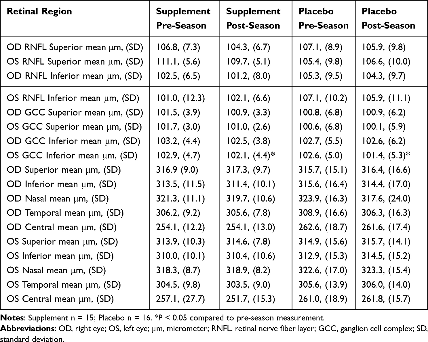
Table 2 Retinal Structure Measurements by Quadrant

Figure 1 Thickness of the Ganglion Cell Complex and Retinal Nerve Fiber Layer inferior and superior regions for both the left (OS); (A–D) and right (OD); (E–H) eyes at pre-season (open circles) and post-season (blue circles) testing in supplement and placebo groups. * p < 0.05 compared to pre-season.
Abbreviations: OS, left eye; OD, right eye; μm, micrometer; GCC, ganglion cell complex; RNFL, retinal nerve fiber layer.
Games played predicted OS central (p = 0.04, R2 =0.133) region thickness with greater thickness at post-season in the placebo group. No other variable in the model predicted thickness.
There were no differences between treatment groups for any structural measurement when using change score (all p > 0.05).
Skin Carotenoid Concentration
Skin carotenoid concentration results are presented in Table 3 and Figure 2. Within groups, skin carotenoid concentration increased at post- vs pre-season in supplement (409.2±76.3 vs 299.3±74.3; p < 0.001, Cohen’s d = 1.46) and placebo groups (323.4±61.3 vs 288.6±51.0, p < 0.001, Cohen’s d = 0.62). Between group comparison showed the supplement group had greater scores at both timepoints, with a significantly higher score at post-season (409.2±76.3 vs 323.4±61.3; p = 0.04, Cohen’s d = 1.24).

Table 3 Skin Carotenoid Concentration
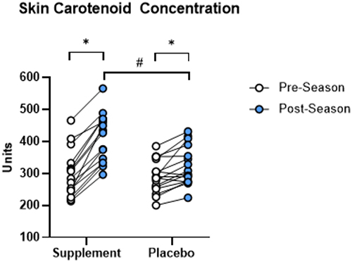
Figure 2 Measurements of total skin carotenoid concentration at pre-season (open circles) and post-season (blue circles) testing in supplement and placebo groups. *p < 0.05 compared to pre-season; # p < 0.05 compared to post-season between groups.
Treatment group predicted skin carotenoid concentration (p = 0.002, R2 =0.292) at post-season.
Change score revealed a difference between treatment groups for skin carotenoid concentration scores (109.9±36.7 vs 34.8±33.0; p < 0.001, Cohen’s d = 2.15) with greater positive change in the supplement group.
Function
Results of visual function are presented in Table 4, and Figures 3 and 4. CFF approached significant difference at post- vs pre-season testing for supplement (27.6±1.8 vs 25.7±2.2 Hz, p = 0.08, Cohen’s d = 0.44) but was not different for the placebo group (26.2±1.8 vs 26.0±1.6 Hz, p = 0.70, Cohen’s d = 0.11). Comparisons between groups were not significant for CFF (p = 0.90, partial η2 =0.001). MPOD was not different at post- vs pre-season for supplement (0.43 ±0.19 vs 0.45±0.21, p = 0.56, Cohen’s d = 0.13) or placebo groups (0.47±0.15 vs 0.40±0.21, p = 0.12, Cohen’s d = 0.37). Between group comparisons were not significant for MPOD (p = 0.99, partial η2 =0.000). No CS trial was different within (p = 0.60, partial η2 =0.228) or between groups (p = 0.59, partial η2 =0.230).
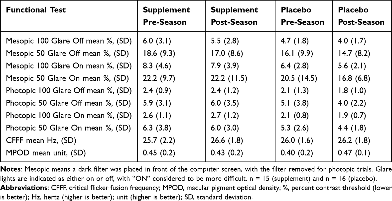
Table 4 Functional Vision Measurements
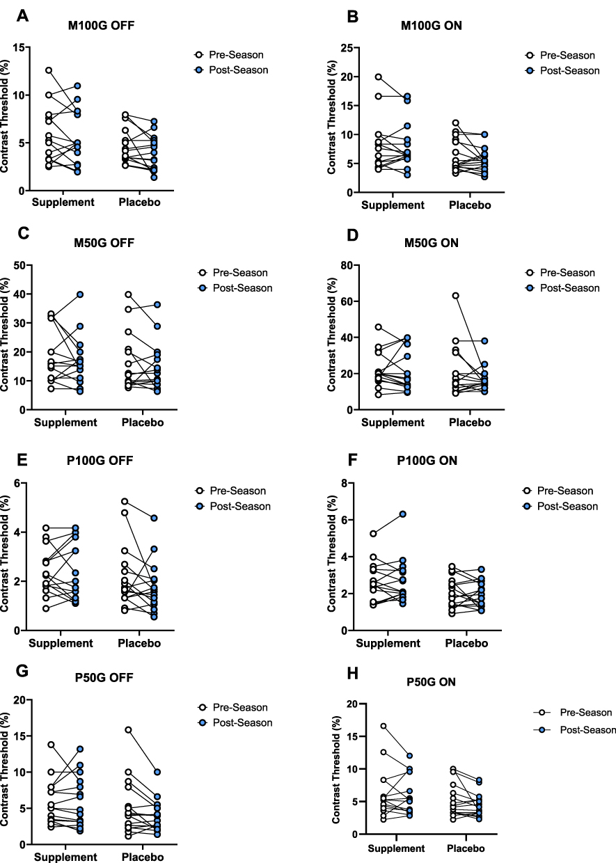
Figure 3 Contrast sensitivity scores for mesopic (A–D) and photopic (E–H) trials at pre-season (open circles) and post-season (blue circles) testing in supplement and placebo groups.
Abbreviations: M100G Off, mesopic-100-glare-off; M100G On, mesopic-100-glare-on; M50G Off, mesopic-50-glare-off; M50G On, mesopic-50-glare-on; P100G Off, photopic-100-glare-off; P100G On, photopic-100-glare-on; P50G Off, photopic-100-glare-off; P50G On, photopic-50-glare-on.
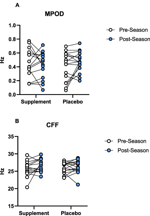
Figure 4 Macular pigment optical density (A) and critical flicker-fusion frequency (B) scores at pre-season (open circles) and post-season (blue circles) testing in supplement and placebo groups.
Abbreviations: MPOD, macular pigment optical density; CFF, critical flicker-fusion frequency.
Concussion history was a predictor of greater MPOD (p = 0.017, R2 =0.182). Concussion history (p = 0.01, R2 =0.205), pills taken (p = 0.025, R2 =0.338), and number of games played (p = 0.027, R2 =0.449) predicted CS trial mesopic-50-glare-off while treatment group predicted mesopic-100-glare-on (p = 0.048, R2 =0.128) and photopic-100-glare-on (p = 0.042, R2 =0.135) trials at post-season. The model did not predict CFF scores (all p > 0.05).
There were no differences between treatment groups for CFF (p = 0.32), MPOD (p = 0.14) or any CS trial (all p > 0.05) when using change score.
Blood Biomarkers
Blood biomarkers are shown in Table 5 and Figure 5. Removal of extreme outliers and those with undetectable concentrations resulted in differing amounts of data included in analysis of NF-L, GFAP, and Tau (14 supplement; 16 placebo), and UCH-L1 (11 supplement; 13 placebo). Using median concentrations and inter quartile range (IQR), NF-L was different at post- vs pre-season in the placebo group (10.05 [7.96–11.48] vs 7.21 [5.57–7.91] pg/mL; p = 0.003) but not the supplement group (7.07 [6.11–13.8] vs 7.78 [5.57–9.75] pg/mL; p = 0.40), with no difference between groups (p = 0.94). GFAP was not different at post- vs pre-season in the supplement (90.73 [78.8–134.63] vs 103 [54.68–125.5] pg/mL; p = 0.94) or placebo groups (101.53 [81.1–114.88] vs 88.78 [63.5–109.5] pg/mL; p = 0.12) with no difference between groups (p = 0.80). Tau was not different at post- vs pre-season in the supplement (1.33 [0.92–1.85] vs 1.2 [0.83–1.39] pg/mL p = 0.31) or placebo groups (1.01 [0.78–1.12] vs 0.87 [0.67–1.12] pg/mL; p = 0.62) but was different between groups at pre- (1.2 [0.83–1.39] vs 0.87 [0.67–1.12] pg/mL; p = 0.04) and post-season (1.33 [0.92–1.85] vs 1.01 [0.78–1.12] pg/mL; p = 0.02) with a greater concentration in the supplement group. UCH-L1 was not different at post- vs pre-season testing for supplement (14.05 [6.97–39.38] vs 16.5 [6.17–33.20] pg/mL; p = 0.90) or placebo groups (8.88 [5.36–17.05] vs 11.22 [6.54–17.55] pg/mL; p = 0.67) with no difference between groups (p = 0.21).
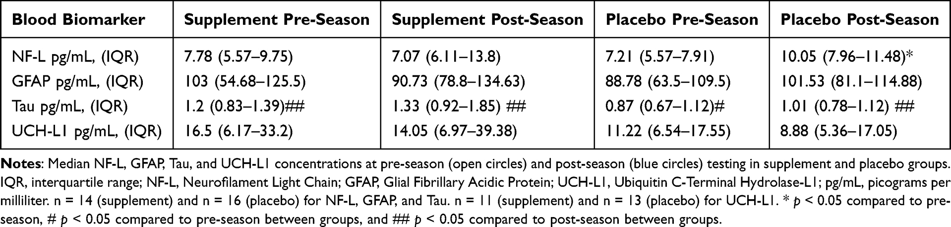
Table 5 Blood Biomarker Concentrations

Figure 5 (A) NF-L, (B) GFAP, (C) Tau, and (D) UCH-L1 concentrations at pre-season (white) and post-season (blue) testing in supplement and placebo groups. Boxes represent first and third quartiles with median values denoted by the horizontal lines, and whiskers indicate minimum and maximum observations. n = 14 (supplement) and n = 16 (placebo) for NF-L, GFAP, and Tau. n = 11 (supplement) and n = 13 (placebo) for UCH-L1. * p < 0.05 compared to pre-season. # p < 0.05 compared to pre-season between groups and ## p < 0.05 compared to post-season between groups.
Abbreviations: NF-L, Neurofilament Light Chain; GFAP, Glial Fibrillary Acidic Protein; UCH-L1, Ubiquitin C-Terminal Hydrolase-L1; pg/mL, picograms per milliliter.
Sex (p = 0.16, R2 =0.204), treatment group (p = 0.006, R2 =0.333), and RHIs per game (p = 0.002, R2 =0.445) predicted Tau concentrations at post-season. The model did not predict NF-L, GFAP, or UCH-L1 concentrations (all p > 0.05).
There were no differences between treatment groups for NF-L (p = 0.16), GFAP (p = 0.40), Tau (p = 0.60), or UCH-L1 (p = 0.40) when using change score (Figure 6).

Figure 6 (A) NF-L, (B) GFAP, (C) Tau, and (D) UCH-L1 change score concentrations from pre-season to post-season in supplement (yellow) and placebo (green) groups. Boxes represent first and third quartiles with median values denoted by the horizontal lines, and whiskers indicate minimum and maximum observations. n = 14 (supplement) and n = 16 (placebo) for NF-L, GFAP, and Tau. n = 11 (supplement) and n = 13 (placebo) for UCH-L1.
Abbreviations: NF-L, Neurofilament Light Chain; GFAP, Glial Fibrillary Acidic Protein; UCH-L1, Ubiquitin C-Terminal Hydrolase-L1; pg/mL, picograms per milliliter.
Discussion
To our knowledge, this study is the first of its kind to implement a supplemental intervention to mitigate damage and associated dysfunction of the visual system following exposure to RHIs. The inferior region of the GCC was thinner at post-season in both groups in the left eye, with more pronounced thinning in the placebo group. Measures of visual function were not different between groups. However, skin carotenoid concentration was greater at post-season in the supplement group. NF-L concentration was elevated in the placebo group at post-season when compared to pre-season, with no significant pre-post season changes in the supplement group. Tau concentration was greater in the supplement group at both time points, significantly at post-season compared to the placebo group. GFAP concentrations were not different within or between groups, though there was a pronounced increase at post-season in the placebo group and decrease in the supplement group. UCH-L1 concentrations were not different within or between for either group. These results and others provide empirical evidence of retinal tissue thinning following exposure to RHIs over the course of one collegiate rugby season. Supplementation with MPCs and EPA + DHA may limit increases in blood biomarkers associated with neurodegeneration and the degree of retinal thinning which in turn would preserve visual function capabilities, but additional research is needed to evaluate these major findings.
Although retinal tissue gradually thins with age, the degree of thinning experienced by participants in this study exceeds accepted norms. GCC thinning in healthy individuals ranges from −0.25 to −0.70 μm/year depending on age.15,75,76 Rugby players experienced an average of −0.80 μm (supplement) and −1.20 μm (placebo) GCC inferior region thinning following one collegiate rugby season from August to November. This represents thinning of 1.5% and 1.7% in the supplement and placebo groups, respectively, vs an expected 0.07% of tissue loss over this time period for this age group. Thinning to the extent experienced by the placebo group is similar to that which glaucoma patients average in one year (−1.18 μm), illustrating the severity of this issue and potential for long-term complications of continued exposure to RHIs.76 Greater preservation of GCC tissue in the supplement group may be attributed to an increased accumulation of lutein, zeaxanthin, meso-zeaxanthin, EPA, and DHA in brain and retinal tissue of participants. The abundance of antioxidative and anti-inflammatory molecules in the supplement group may have acted as a defense mechanism from oxidative stress and chronic inflammation resulting from RHIs, seen previously in rodent head injury models.47,51–54 The presence of MPCs and omega 3’s would allow for the donation of electrons to stabilize free radicals and combat sustained inflammation while preserving neuronal integrity.77,78 Thinning of the inferior region is expected prior to the detection of superior thinning due to anatomical vulnerability.79 While interesting, thinning of only left eye tissue is not unique as it has been previously observed in retired rugby players.80 Further, asymmetry of tissue thickness between eyes has been observed in multiple collision sports, including rugby players in our previous study (under review).16,17 This may be simply explained by localized neuroinflammation from hits to the left side of participants caused by learned tackling and avoidance techniques but is beyond the scope of this study.81 Surprisingly, more games played predicted greater OS central thickness. However, this finding is similar to previous work concerning thickening of retinal tissue in football players, requiring continued research to determine an explanation.82 Despite an absence of thinning in RNFL or RM regions, GCC thinning is believed to precede these regions.83 As such, RNFL thinning in rugby players is expected to take place following continued participation as seen in those with an average of 18 years of participation.80 Thus, a longitudinal design is proposed to illuminate such thinning, as evidenced by previous research concerning collision sport athletes.16,17
CFF, MPOD, and CS were not different, but CFF did increase more in the supplement group at post-season evaluation. Although not a statistically significant change in CFF, the increase in the supplement group is indicative of a trending change observed in prior studies performed over slightly longer timelines.84 Our CFF, MPOD, and CS scores at baseline and follow-up are supported by Stringham et al72 who found CFF increases in both supplement and placebo groups at 3-month follow-up. Further, MPOD and CS were not different at 3-month follow-up with varying trends in score-change direction, mirroring our results.72 Moreover, greatest improvements in CFF were seen at 6-month follow-up.72 Participants in the present study completed CFF testing at the midway point of the testing battery in a rested state. As such, a non-significant increase in score for the supplement group may be attributed to the conditions in which the test was taken. Indeed, a rested state may have resulted in altered cognitive processing, but perhaps the benefits of supplementation present themselves during rugby participation.85 Interestingly, those with a history of concussion were predicted to have higher MPOD. Dietary interventions are often prescribed following concussive injury, conceivably resulting in a lifestyle change leading to a natural increase in MPOD in these participants over time.86 Variation in MPOD score supports both a longer duration study and improved methodology of measuring supplement compliance. Our findings are supported by Nolan and colleagues87 who observed increases in MPOD predominantly at 12-month follow-up, but not at 3 months. Further, increases in MPOD were not associated with CS improvement in those supplementing with MPCs.87 Association between MPOD and CS is disputed, with a weak relation even when supplementing with MPCs or analyzing dietary intake.68,88,89 Conversely, no association between MPOD and CS in a population of adults aged 20–3590 and 18–4091 was reported in various studies. No difference between CS scores for our groups contradicts the work of Leong et al,16 though athletes in that sample were significantly older and thus likely to have been exposed to RHIs for a greater amount of time compared to our athletes (19 vs 46 years old).16 Moreover, functional deficits are not expected until 25–35% of GCC tissue atrophies, meaning athletes from our sample may experience functional deficits to a similar degree as those in the sample of Leong et al16 if thinning persists at this rate.15,16 More concussions and games played predicted worse CS performance on the mesopic-50-glare-off trial while number of pills taken predicted an improvement on this trial, all expected findings. Unexpectedly, treatment group predicted better scores on mesopic-100-glare-on and photopic-100-glare-on with the placebo group performing better, potentially explained by the short duration of supplementation. Examination of the literature supports supplementation with MPCs for more than 3-months to elicit significant CFF, MPOD, and CS improvement. Actively engaging in consumption of MPCs through diet or supplementation appears beneficial for increasing MPOD and in-turn improving visual function.80,92 Further, omega-3 consumption is often promoted to patients to improve ocular health, supporting a combination of MPCs + omega-3s.62
Skin carotenoid concentration improved in both groups, with a greater level at post-season for the supplement group compared with the placebo group. Unsurprisingly, treatment group predicted skin carotenoid concentration at post-season. Improvement of this magnitude is crucial for substantiating test supplement bioavailability. While unexpected that the placebo group significantly increased from pre- to post-season, this may be explained by conscious dietary manipulation to meet the nutritional needs of a competition season.93 Mean scores for each group are consistent with previous work for supplementation status and duration of supplementation,94 and indicate strong compliance to supplement ingestion. Significant increases in skin carotenoid levels over three months and beyond are well documented following supplementation with MPCs95–97 and imply that the supplement was successful in reaching target tissue, a strong correlate with retinal98 and cerebral99 concentrations. Beyond the known visual benefits of increased skin carotenoid concentration, resistance to ultraviolet radiation is another advantage.100 Although increases in skin concentration tend to level-off, continued supplementation and dietary intake is required to maintain the plethora of benefits provided.92,94
Elevated concentrations of NF-L are well established in neurodegenerative populations including Alzheimer’s disease, amyotrophic lateral sclerosis, and Parkinson’s disease.101,102 Importantly, an increase in NF-L has been associated with thinner GCC tissue in individuals with neurological dysfunction.103 Our results support this finding as the placebo group experienced significantly elevated NF-L and greater GCC thinning after one collegiate rugby season while the supplement group experienced GCC thinning to a lesser extent and lower NF-L concentrations. These findings suggest macular pigment carotenoid + omega-3 supplementation may attenuate the deleterious effects of RHIs throughout a rugby season by preserving retinal tissue and neuronal integrity. Although not significant, GFAP concentrations in the supplement group were lower at post-season while the placebo group saw a large increase. Though speculative, this may be due to the supplement preserving the integrity of astroglial cytoskeletons, while the placebo group remained susceptible to damage from RHIs. Rodent models subjected to brain injury and subsequently treated with lutein and zeaxanthin show decreased GFAP concentrations compared to non-treatment groups, supporting the downward trend of our findings and warranting continued exploration.51 Together, both NF-L and GFAP concentrations saw downward trends in the supplement group while the placebo group experienced upward trends conveying both the danger of exposure to RHIs and possible effectiveness of the test supplement. This is particularly notable given the greater rate of RHIs experienced by the supplement group, and as such, further work on this finding may be a primary focus in future related research. As Tau is elevated following exposure to RHIs and in retired rugby players with a history of concussion,104,105 an increase in Tau was expected. Though both groups saw an increase at post-season, the supplement group had a higher concentration compared to the placebo group which was unexpected. This may be explained by imperfect Tau-targeting abilities of the supplement and the heterogeneity of human Tau pathologies, both known hurdles to treating Tau accumulations.106 It may also reflect the performance of the assay itself as new Tau assays are being developed. Further, sex (women) and more RHIs per game predicted higher Tau concentrations, of which there were more of both in the supplement group. Additionally, the supplement group played significantly more minutes per game, theoretically exposing them to more RHIs. Thus, exposure to more RHIs per game and the complexity of therapeutically targeting Tau may explain the different concentrations at post-season. UCH-L1 has shown promise as an acute biomarker of injury.31 However, due to its short half-life (< 12 hours), it is challenging to detect long-term changes which likely explains the lack of difference within and between groups in the current study.107–109 As longer supplementation periods have proven beneficial,68,110 future research must evaluate a period greater than 3 months to gain a better understanding of the promising trends seen here while consuming the test supplement and utilizing a larger sample size to prevent Type II error.111
Limitations
Limitations to the present study include no quantification of RHIs, self-reported compliance, a short duration of supplementation, a relatively small sample size, pupil size measure, and imperfect CS measures. Incorporation of technology to objectively quantify RHIs experienced by participants will strengthen results. Although impractical, observing daily supplement intake would improve compliance rates and eliminate a reliance on participants to practice self-intake. However, this is not realistic in daily life as humans are prone to forgetfulness, especially when undertaking a lifestyle change. Next, a longer period of supplementation to accurately quantify outcome measures is supported in the literature. Nevertheless, this study encapsulates the true duration of a collegiate rugby season, representing an opportunity to explore supplemental outcomes of a shorter duration not often found in the literature. Additionally, a larger sample size is required from which conclusions may be drawn. While appropriately powered for visual analysis, blood biomarker analysis often requires a larger sample size. Also, objective measure of pupil size at time of scan would increase reliability of CFF measures. Finally, it should be noted that some argument has been posed as to the reliability of monitor-based based CS112 measures, thus, future studies may consider using alternative optical systems for CS.
Conclusion
Collegiate rugby players demonstrate GCC thinning and increased concentrations of NF-L and Tau following one season. A supplemental intervention of lutein, zeaxanthin, meso-zeaxanthin, and EPA + DHA may be effective in limiting the extent of tissue loss in these athletes while also preventing a significant increase in NF-L, whereas its ability to limit increases in Tau requires further study and evaluation with newer assays. Persistent, accelerated thinning of retinal tissue will result in visual dysfunction if not acted upon. Though visual function was not improved, supplementation resulted in a rapid increase in skin carotenoid concentration in just three months, confirming exceptional bioavailability of the test supplement and suggesting potential CFF, MPOD, and CS improvement following a longer supplementation period. This study is important for broadening the understanding of damage to the visual system following exposure to RHIs and has significant implications for prevention and treatment opportunities of associated damage through supplemental intervention.
Data Sharing Statement
Individual deidentified participant data, including OCT, visual function scores, skin carotenoid concentration, and inflammatory blood serum biomarker concentration, may be made available upon reasonable request to the corresponding author. Study consent form may also be made available upon exceptional request to the corresponding author. Accepted data sharing requests will be opened for an agreed upon time period between the requesting party and the authorship team.
Acknowledgments
The authors would like to thank Cyndi Flanagan and Christa Oelhaf of the Penn State Clinical Research Center for their assistance with biological sample collection, as well as Harvey Hanlen and members of the Penn State Sport Concussion Research and Service Lab for their support.
Funding
The authors received financial support from MacuHealth LLC. The sponsor was not involved in study design, recruitment, data collection, or data analysis.
Disclosure
James Stringham receives a salary from MacuHealth LLC where he is the chief scientific officer. In addition, Dr James Stringham has a patent Nutritional Supplement to Reduce Severity of and Enhance Recovery From Repetitive Head Impacts pending to MacuHealth LLC. The authors received financial support from MacuHealth LLC. The sponsor was not involved in study design, recruitment, data collection, or data analysis. The authors report no other conflicts of interest in this work.
References
1. Stewart W, Buckland ME, Abdolmohammadi B, et al. Risk of chronic traumatic encephalopathy in rugby union is associated with length of playing career. Acta Neuropathol. 2023;146(6):829. doi:10.1007/s00401-023-02644-3
2. McKee AC, Mez J, Abdolmohammadi B, et al. Neuropathologic and clinical findings in young contact sport athletes exposed to repetitive head impacts. JAMA Neurol. 2023;80(10):1037–1050. doi:10.1001/jamaneurol.2023.2907
3. McKee AC, Stein TD, Huber BR, et al. Chronic traumatic encephalopathy (CTE): criteria for neuropathological diagnosis and relationship to repetitive head impacts. Acta Neuropathol. 2023;145(4):371–394. doi:10.1007/s00401-023-02540-w
4. Scorza KA, Cole W. Current concepts in concussion: initial evaluation and management. Am Family Phys. 2019;99(7):426–434.
5. Tierney GJ, Simms CK. Can tackle height influence head injury assessment risk in elite rugby union? J Sci Med Sport. 2018;21(12):1210–1214. doi:10.1016/j.jsams.2018.05.010
6. Tucker R, Raftery M, Kemp S, et al. Risk factors for head injury events in professional rugby union: a video analysis of 464 head injury events to inform proposed injury prevention strategies. Br J Sports Med. 2017;51(15):1152–1157. doi:10.1136/bjsports-2017-097895
7. Woodward J, Tooby J, Tucker R, et al. Instrumented mouthguards in elite-level men’s and women’s rugby union: characterising tackle-based head acceleration events. BMJ Open Sport Exercise Med. 2024;10(3):e002013. doi:10.1136/bmjsem-2024-002013
8. Kawasaki T, Kawakami Y, Nojiri S, et al. Risk factors for concussion in under 18, under 22 and professional men’s rugby union: a video analysis of 14,809 tackles. Sports Med – Open. 2023;9(1):95. doi:10.1186/s40798-023-00642-z
9. Tooby J, Weaving D, Al-Dawoud M, et al. Quantification of head acceleration events in rugby league: an Instrumented mouthguard and video analysis pilot study. Sensors. 2022;22(2):584. doi:10.3390/s22020584
10. King D, Hume P, Gissane C, et al. Measurement of head impacts in a senior amateur rugby league team with an instrumented patch: exploratory analysis. ARC J Res Sports Med. 2017;2.
11. Peek RJ, Carey DL, Middleton KJ, et al. Association between position-specific impact and movement characteristics of professional rugby union Players during game play. J Strength Conditioning Res. 2023;37(1):161–166. doi:10.1519/JSC.0000000000004170
12. Datta S, Baidya K, Banerjee M, et al. Retinal nerve fibre layer thinning in patients with thalassaemia, iron deficiency anaemia, and anaemia of chronic diseases. J Ophthalmol. 2020;2020:9268364. doi:10.1155/2020/9268364
13. Kashani AH, Zimmer-Galler IE, Shah SM, et al. Retinal thickness analysis by race, gender, and age using stratus OCT™. Am J Ophthalmol. 2010;149(3):496–502.e1. doi:10.1016/j.ajo.2009.09.025
14. Stepp MA, Menko AS. Immune responses to injury and their links to eye disease. Transl Res. 2021;236:52. doi:10.1016/j.trsl.2021.05.005
15. Scuderi G, Fragiotta S, Scuderi L, et al. Ganglion cell complex analysis in glaucoma patients: what can it tell us? Eye and Brain. 2020;12:33–44. doi:10.2147/EB.S226319
16. Leong D, Morettin C, Messner LV, et al. Visual structure and function in collision sport athletes. J Neuroophthalmol. 2018;38(3):285–291. doi:10.1097/WNO.0000000000000572
17. Childs C, Barker LA, Gage AM, et al. Investigating possible retinal biomarkers of head trauma in Olympic boxers using optical coherence tomography. Eye and Brain. 2018;10:101. doi:10.2147/EB.S183042
18. Schrupp LE, Ciuffreda KJ, Kapoor N. Foveal versus eccentric retinal critical flicker frequency in mild traumatic brain injury. Optometry. 2009;80(11):642–650. doi:10.1016/j.optm.2009.04.097
19. García-Romera M-C, Silva-Viguera M-C, López-Izquierdo I, et al. Effect of macular pigment carotenoids on cognitive functions: a systematic review. Physiol Behav. 2022;254:113891. doi:10.1016/j.physbeh.2022.113891
20. Kawata K, Rubin LH, Lee JH, et al. Association of football subconcussive head impacts with ocular near point of convergence. JAMA Ophthalmol. 2016;134(7):763–769. doi:10.1001/jamaophthalmol.2016.1085
21. Zonner SW, Ejima K, Fulgar CC, et al. Oculomotor response to cumulative subconcussive head impacts in US High School Football Players: a pilot longitudinal study. JAMA Ophthalmol. 2019;137(3):265–270. doi:10.1001/jamaophthalmol.2018.6193
22. Nowak MK, Bevilacqua ZW, Ejima K, et al. Neuro-ophthalmologic response to repetitive subconcussive head impacts. JAMA Ophthalmol. 2020;138(4):350–357. doi:10.1001/jamaophthalmol.2019.6128
23. Johnson B, Zhang K, Hallett M, et al. Functional neuroimaging of acute oculomotor deficits in concussed athletes. Brain Imaging Behav. 2015;9(3):564–573. doi:10.1007/s11682-014-9316-x
24. Johnson B, Hallett M, Slobounov S. Follow-up evaluation of oculomotor performance with fMRI in the subacute phase of concussion. Neurology. 2015;85(13):1163. doi:10.1212/WNL.0000000000001968
25. Soriano S, Curry K, Sadrameli SS, et al. Alterations to the gut microbiome after sport-related concussion in a collegiate football players cohort: a pilot study. Brain Behav Immun- Health. 2022;21:100438. doi:10.1016/j.bbih.2022.100438
26. Papa L, Walter AE, Wilkes JR, et al. Effect of player position on serum biomarkers during participation in a season of collegiate football. J Neurotrauma. 2022;39(19–20):1339–1348. doi:10.1089/neu.2022.0083
27. Asken BM, Tanner JA, VandeVrede L, et al. Multi-modal biomarkers of repetitive head impacts and traumatic encephalopathy syndrome: a clinicopathological case series. J Neurotrauma. 2022;39(17–18):1195–1213. doi:10.1089/neu.2022.0060
28. Bernick C, Shan G, Ritter A, et al. Blood biomarkers and neurodegeneration in individuals exposed to repetitive head impacts. Alzheimer’s Res Ther. 2023;15:173. doi:10.1186/s13195-023-01310-w
29. Joseph JR, Swallow JS, Willsey K, et al. Elevated markers of brain injury as a result of clinically asymptomatic high-acceleration head impacts in high-school football athletes. J Neurosurg. 2019;130(5):1642–1648. doi:10.3171/2017.12.JNS172386
30. Puvenna V, Brennan C, Shaw G, et al. Significance of ubiquitin carboxy-terminal hydrolase L1 elevations in athletes after sub-concussive head hits. PLoS One. 2014;9(5):e96296. doi:10.1371/journal.pone.0096296
31. Lember L-M, Ntikas M, Mondello S, et al. The use of biofluid markers to evaluate the consequences of sport-related subconcussive head impact exposure: a scoping review. Sports Med – Open. 2024;10:12. doi:10.1186/s40798-023-00665-6
32. Wang KK, Yang Z, Zhu T, et al. An update on diagnostic and prognostic biomarkers for traumatic brain injury. Expert Rev Molecular Diagnostics. 2018;18(2):165. doi:10.1080/14737159.2018.1428089
33. Miner AE, Groh JR, Tripodis Y, et al. Examination of plasma biomarkers of amyloid, tau, neurodegeneration, and neuroinflammation in former elite American football players. Alzheimer’s Dementia. 2024;20(11):7529. doi:10.1002/alz.14231
34. Tabor JB, Penner LC, Galarneau J-M, et al. Plasma biomarkers of traumatic brain injury in adolescents with sport-related concussion. JAMA Network Open. 2024;7(9):e2431959. doi:10.1001/jamanetworkopen.2024.31959
35. Mares J. Lutein and zeaxanthin isomers in eye health and disease. Ann Rev Nutr. 2016;36:571–602. doi:10.1146/annurev-nutr-071715-051110
36. Arunkumar R, Gorusupudi A, Bernstein PS. The macular carotenoids: a biochemical overview. Biochimica et biophysica acta Mol Cell Biol Lipids. 2020;1865(11):158617. doi:10.1016/j.bbalip.2020.158617
37. Demmig-Adams B, López-Pozo M, Stewart JJ, et al. Zeaxanthin and lutein: photoprotectors, anti-inflammatories, and brain food. Molecules. 2020;25(16):3607. doi:10.3390/molecules25163607
38. Arunkumar R, Calvo CM, Conrady CD, et al. What do we know about the macular pigment in AMD: the past, the present, and the future. Eye. 2018;32(5):992–1004. doi:10.1038/s41433-018-0044-0
39. Stringham JM, Hammond BR. Macular pigment and visual performance under glare conditions. Optometry Vision Sci. 2008;85(2):82–88. doi:10.1097/OPX.0b013e318162266e
40. Stringham JM, O’Brien KJ, Stringham NT. Macular carotenoid supplementation improves disability glare performance and dynamics of photostress recovery. Eye and Vision. 2016;3:30. doi:10.1186/s40662-016-0060-8
41. Stringham JM, Johnson EJ, Hammond BR. Lutein across the lifespan: from childhood cognitive performance to the aging eye and brain. Curr Dev Nutr. 2019;3(7):nzz066. doi:10.1093/cdn/nzz066
42. Kuse Y, Tsuruma K, Kanno Y, et al. CCR3 is associated with the death of a photoreceptor cell-line induced by light exposure. Front Pharmacol. 2017;8:–2017. doi:10.3389/fphar.2017.00207
43. Chen P, Lin A. Tectonic topography and late pleistocene activity of the West Qinling Fault, northeastern Tibetan Plateau. J Asian Earth Sci. 2019;176:68–78. doi:10.1016/j.jseaes.2019.02.007
44. Sparrow JR, Cai B. Blue Light–Induced Apoptosis of A2E-containing RPE: involvement of Caspase-3 and protection by Bcl-2. Invest Ophthalmol Visual Sci. 2001;42(6):1356–1362.
45. Sparrow JR, Zhou J, Cai B. DNA is a target of the photodynamic effects elicited in A2E-Laden RPE by blue-light illumination. Invest Ophthalmol Visual Sci. 2003;44(5):2245–2251. doi:10.1167/iovs.02-0746
46. Bernstein PS, Delori FC, Richer S, et al. The value of measurement of macular carotenoid pigment optical densities and distributions in age-related macular degeneration and other retinal disorders. Vision Res. 2010;50(7):716–728. doi:10.1016/j.visres.2009.10.014
47. Sasaki M, Ozawa Y, Kurihara T, et al. Neuroprotective effect of an antioxidant, lutein, during retinal inflammation. Invest Ophthalmol Visual Sci. 2009;50(3):1433–1439. doi:10.1167/iovs.08-2493
48. Ozawa Y, Sasaki M, Takahashi N, et al. Neuroprotective effects of lutein in the retina. Curr Pharm Des. 2012;18(1):51–56. doi:10.2174/138161212798919101
49. Stringham NT, Holmes PV, Stringham JM. Effects of macular xanthophyll supplementation on brain-derived neurotrophic factor, pro-inflammatory cytokines, and cognitive performance. Physiol Behav. 2019;211:112650. doi:10.1016/j.physbeh.2019.112650
50. Kelly J, Griffith O, Papa L, et al. Perspective: supplementation with macular pigment carotenoids with respect to brain injury. Discover Medicine. 2025;2(1):1–9. doi:10.1007/s44337-025-00190-3
51. Gunal MY, Sakul AA, Caglayan AB, et al. Protective effect of lutein/zeaxanthin isomers in traumatic brain injury in mice. Neurotox Res. 2021;39(5):1543–1550. doi:10.1007/s12640-021-00385-3
52. Tan D, Yu X, Chen M, et al. Lutein protects against severe traumatic brain injury through anti‑inflammation and antioxidative effects via ICAM‑1/Nrf‑2. Mol Med Rep. 2017;16(4):4235–4240. doi:10.3892/mmr.2017.7040
53. Sun YX, Liu T, Dai X-L, et al. Treatment with lutein provides neuroprotection in mice subjected to transient cerebral ischemia. J Asian Nat Prod Res. 2014;16(11):1084–1093. doi:10.1080/10286020.2014.939584
54. Turk C, Camlar M, Diniz G, et al. Effects of lutein on brain damage and vasospasm in an experimental subarachnoid hemorrhage model. World Neurosurg. 2020;143:e450–e455. doi:10.1016/j.wneu.2020.07.186
55. Jeromson S, Gallagher I, Galloway S, et al. Omega-3 fatty acids and skeletal muscle health. Mar Drugs. 2015;13(11):6977. doi:10.3390/md13116977
56. Troesch B, Eggersdorfer M, Laviano A, et al. Expert opinion on benefits of long-chain omega-3 fatty acids (DHA and EPA) in aging and clinical nutrition. Nutrients. 2020;12(9):2555. doi:10.3390/nu12092555
57. Kiecolt-Glaser JK, Belury MA, Andridge R, et al. Omega-3 supplementation lowers inflammation and anxiety in medical students: a randomized controlled trial. Brain Behav Immun. 2011;25(8):1725. doi:10.1016/j.bbi.2011.07.229
58. Armstrong A, Anzalone AJ, Pethick W, et al. An evaluation of omega-3 status and intake in Canadian Elite Rugby 7s Players. Nutrients. 2021;13(11):3777. doi:10.3390/nu13113777
59. Djuricic I, Calder PC. Beneficial outcomes of omega-6 and omega-3 polyunsaturated fatty acids on human health: an update for 2021. Nutrients. 2021;13(7):2421. doi:10.3390/nu13072421
60. Heshmati J, Morvaridzadeh M, Maroufizadeh S, et al. Omega-3 fatty acids supplementation and oxidative stress parameters: a systematic review and meta-analysis of clinical trials. Pharmacol Res. 2019;149:104462. doi:10.1016/j.phrs.2019.104462
61. Hajianfar H, Paknahad Z, Bahonar A. The effect of omega-3 supplements on antioxidant capacity in patients with type 2 diabetes. Int J Preventive Med. 2013;4(Suppl 2):S234–238.
62. Zhang AC, Singh S, Craig JP, et al. Omega-3 fatty acids and eye health: opinions and self-reported practice behaviors of optometrists in Australia and New Zealand. Nutrients. 2020;12(4).
63. Johnson EJ, Mcdonald K, Caldarella SM, et al. Cognitive findings of an exploratory trial of docosahexaenoic acid and lutein supplementation in older women. Nutr Neurosci. 2008;11(2):75–83. doi:10.1179/147683008X301450
64. Begtrup KM, Krag AE, Hvas AM. No impact of fish oil supplements on bleeding risk: a systematic review. Dan Med J. 2017;64(5).
65. Maroon JC, Bost JW. ω-3 Fatty acids (fish oil) as an anti-inflammatory: an alternative to nonsteroidal anti-inflammatory drugs for discogenic pain. Surg Neurol. 2006;65(4):326–331. doi:10.1016/j.surneu.2005.10.023
66. Mazza M, Pomponi M, Janiri L, et al. Omega-3 fatty acids and antioxidants in neurological and psychiatric diseases: an overview. Prog Neuro Psychopharmacol Biol Psychiatry. 2007;31(1):12–26. doi:10.1016/j.pnpbp.2006.07.010
67. Zwilling CE, Strang A, Anderson E, et al. Enhanced physical and cognitive performance in active duty Airmen: evidence from a randomized multimodal physical fitness and nutritional intervention. Sci Rep. 2020;10(1):17826. doi:10.1038/s41598-020-74140-7
68. Stringham JM, O’Brien KJ, Stringham NT. Contrast sensitivity and lateral inhibition are enhanced with macular carotenoid supplementation. Invest Ophthalmol Visual Sci. 2017;58(4):2291–2295. doi:10.1167/iovs.16-21087
69. Opitz DL, Tung S, Jang US, et al. Silicone punctal plugs as an adjunctive therapy for open‐angle glaucoma and ocular hypertension. Clin Exp Optometry. 2011;94(5):438–442. doi:10.1111/j.1444-0938.2010.00587.x
70. Chandrakumar M, Colpa L, Reginald YA, et al. Measuring contrast sensitivity using the M&S smart system II versus the Pelli-Robson Chart. Ophthalmology. 2013;120(10):2160–2161.e1. doi:10.1016/j.ophtha.2013.07.022
71. Wooten BR, Hammond BR, Land RI, et al. A practical method for measuring macular pigment optical density. Invest Ophthalmol Vis Sci. 1999;40(11):2481–2489.
72. Stringham JM, Stringham NT, O’Brien KJ. Macular carotenoid supplementation improves visual performance, sleep quality, and adverse physical symptoms in those with high screen time exposure. Foods. 2017;6(7):47. doi:10.3390/foods6070047
73. Hammond BR, Wooten BR, Snodderly DM. Individual variations in the spatial profile of human macular pigment. J Optical Soc Am. 1997;14(6):1187–1196. doi:10.1364/JOSAA.14.001187
74. Stringham JM, Stringham NT. Serum and retinal responses to three different doses of macular carotenoids over 12 weeks of supplementation. Exp Eye Res. 2016;151:1–8. doi:10.1016/j.exer.2016.07.005
75. Zhang X, Francis BA, Dastiridou A, et al. Longitudinal and cross-sectional analyses of age effects on retinal nerve fiber layer and ganglion cell complex thickness by fourier-domain OCT. Trans Vision Sci Technol. 2016;5(2):1.
76. Hou H, Moghimi S, Proudfoot JA, et al. Ganglion cell complex thickness and macula vessel density loss in primary open angle glaucoma. Ophthalmology. 2020;127(8):1043–1052. doi:10.1016/j.ophtha.2019.12.030
77. Li B, Ahmed F, Bernstein PS. Studies on the singlet oxygen scavenging mechanism of human macular pigment. Arch Biochem Biophys. 2010;504(1):56–60. doi:10.1016/j.abb.2010.07.024
78. Anderson EJ, Thayne KA, Harris M, et al. Do fish oil omega-3 fatty acids enhance antioxidant capacity and mitochondrial fatty acid oxidation in human atrial myocardium via PPARγ activation? Antioxid Redox Signal. 2014;21(8):1156. doi:10.1089/ars.2014.5888
79. Ghita AM, Iliescu DA, Ghita AC, et al. Ganglion cell complex analysis: correlations with retinal nerve fiber layer on optical coherence tomography. Diagnostics. 2023;13(2):266. doi:10.3390/diagnostics13020266
80. Kelman JC, Hodge C, Stanwell P, et al. Retinal nerve fibre changes in sports-related repetitive traumatic brain injury. Clin Exp Ophthalmol. 2020;48(2):204–211. doi:10.1111/ceo.13673
81. Sobue S, Kawasaki T, Hasegawa Y, et al. Tackler’s head position relative to the ball carrier is highly correlated with head and neck injuries in rugby. Br J Sports Med. 2017;52(6):353. doi:10.1136/bjsports-2017-098135
82. Silverstein SM, Atlas J, Young M, et al. Football-related concussions and head impacts are associated with changes in retinal structure and signaling. Biomarkers Neuropsychiatry. 2024;10:100091. doi:10.1016/j.bionps.2024.100091
83. Tieger MG, Hedges TR, Ho J, et al. Ganglion cell complex loss in chiasmal compression by brain tumors. J Neuro-Ophthalmol. 2017;37(1):7–12. doi:10.1097/WNO.0000000000000424
84. Bovier ER, Renzi LM, Hammond BR. A double-blind, placebo-controlled study on the effects of lutein and zeaxanthin on neural processing speed and efficiency. PLoS One. 2014;9(9):e108178. doi:10.1371/journal.pone.0108178
85. Maciejewska K, Greń A, Wieczorek A. The effect of acute, moderate intensity indoor cycling on the temporal resolution of human vision system, measured by critical fusion frequency. Physiological Reports. 2020;8(21):e14618. doi:10.14814/phy2.14618
86. Conti F, McCue JJ, DiTuro P, et al. Mitigating traumatic brain injury: a narrative review of supplementation and dietary protocols. Nutrients. 2024;16(15):2430. doi:10.3390/nu16152430
87. Nolan JM, Loughman J, Akkali MC, et al. The impact of macular pigment augmentation on visual performance in normal subjects: COMPASS. Vision Res. 2011;51(5):459–469. doi:10.1016/j.visres.2010.12.016
88. Loughman J, Akkali MC, Beatty S, et al. The relationship between macular pigment and visual performance. Vision Res. 2010;50(13):1249–1256. doi:10.1016/j.visres.2010.04.009
89. Renzi LM, Hammond BR. The effect of macular pigment on heterochromatic luminance contrast. Exp Eye Res. 2010;91(6):896–900. doi:10.1016/j.exer.2010.09.015
90. Estévez-Santiago R, Olmedilla-Alonso B, Beltrán-de-miguel B. Assessment of lutein and zeaxanthin status and dietary markers as predictors of the contrast threshold in 2 age groups of men and women. Nutr Res. 2016;36(7):719–730. doi:10.1016/j.nutres.2016.02.007
91. Kvansakul J, Rodriguez‐Carmona M, Edgar DF, et al. Supplementation with the carotenoids lutein or zeaxanthin improves human visual performance. Ophthalmic Physiol Opt. 2006;26(4):362–371. doi:10.1111/j.1475-1313.2006.00387.x
92. Johnson EJ, Avendano EE, Mohn ES, et al. The association between macular pigment optical density and visual function outcomes: a systematic review and meta-analysis. Eye. 2020;35(6):1620. doi:10.1038/s41433-020-01124-2
93. Jenner S, Belski R, Devlin B, et al. A qualitative investigation of factors influencing the dietary intakes of professional Australian football players. Int J Environ Res Public Health. 2021;18(8):4205. doi:10.3390/ijerph18084205
94. Ermakov IV, Ermakova M, Sharifzadeh M, et al. Optical assessment of skin carotenoid status as a biomarker of vegetable and fruit intake. Arch Biochem Biophys. 2018;646:46–54. doi:10.1016/j.abb.2018.03.033
95. Palombo P, Fabrizi G, Ruocco V, et al. Beneficial long-term effects of combined oral/topical antioxidant treatment with the carotenoids lutein and zeaxanthin on human skin: a double-blind, placebo-controlled study. Skin Pharmacol Physiol. 2007;20(4):199–210. doi:10.1159/000101807
96. Baswan SM, Marini A, Klosner AE, et al. Orally administered mixed carotenoids protect human skin against ultraviolet A-induced skin pigmentation: a double-blind, placebo-controlled, randomized clinical trial. Photodermatol Photoimmunol Photomed. 2020;36(3):219–225. doi:10.1111/phpp.12541
97. Obana A, Gohto Y, Nakazawa R, et al. Effect of an antioxidant supplement containing high dose lutein and zeaxanthin on macular pigment and skin carotenoid levels. Sci Rep. 2020;10:10262. doi:10.1038/s41598-020-66962-2
98. Conrady CD, Bell JP, Besch BM, et al. Correlations between macular, skin, and serum carotenoids. Invest Ophthalmol Visual Sci. 2017;58(9):3616–3627. doi:10.1167/iovs.17-21818
99. Vishwanathan R, Neuringer M, Snodderly DM, et al. Macular lutein and zeaxanthin are related to brain lutein and zeaxanthin in primates. Nutr Neurosci. 2013;16(1):21–29. doi:10.1179/1476830512Y.0000000024
100. Baswan SM, Klosner AE, Weir C, et al. Role of ingestible carotenoids in skin protection: a review of clinical evidence. Photodermatol Photoimmunol Photomed. 2021;37(6):490–504. doi:10.1111/phpp.12690
101. Gaiottino J, Norgren N, Dobson R, et al. Increased neurofilament light chain blood levels in neurodegenerative neurological diseases. PLoS One. 2013;8(9):e75091. doi:10.1371/journal.pone.0075091
102. Buhmann C, Magnus T, Choe C-U. Blood neurofilament light chain in Parkinson’s disease. J Neural Transm. 2023;130(6):755. doi:10.1007/s00702-023-02632-7
103. Garzone D, Finger RP, Mauschitz MM, et al. Neurofilament light chain and retinal layers’ determinants and association: a population‐based study. Ann Clin Transl Neurol. 2022;9(4):564. doi:10.1002/acn3.51522
104. Cente M, Perackova J, Peracek P, et al. Association of nonconcussive repetitive head impacts and intense physical activity with levels of phosphorylated Tau 181 and total Tau in plasma of young elite soccer players. JAMA Network Open. 2023;6(3):e236101. doi:10.1001/jamanetworkopen.2023.6101
105. Alanazi N, Fitzgerald M, Hume P, et al. Concussion-related biomarker variations in retired rugby players and implications for neurodegenerative disease risk: the UK Rugby Health Study. Int J Mol Sci. 2024;25(14):7811. doi:10.3390/ijms25147811
106. Samudra N, Lane-Donovan C, VandeVrede L, et al. Tau pathology in neurodegenerative disease: disease mechanisms and therapeutic avenues. J Clin Invest. 2023;133(12):e168553. doi:10.1172/JCI168553
107. Papa L, Brophy GM, Welch RD, et al. Time course and diagnostic accuracy of glial and neuronal blood biomarkers GFAP and UCH-L1 in a large cohort of trauma patients with and without mild traumatic brain injury. JAMA Neurol. 2016;73(5):551. doi:10.1001/jamaneurol.2016.0039
108. Korley FK, Jain S, Sun X, et al. Prognostic value of day-of-injury plasma GFAP and UCH-L1 levels for predicting functional recovery in the TRACK-TBI cohort: an observational cohort study. Lancet Neurol. 2022;21(9):803. doi:10.1016/S1474-4422(22)00256-3
109. Brophy GM, Mondello S, Papa L, et al. Biokinetic analysis of Ubiquitin C-Terminal Hydrolase-L1 (UCH-L1) in severe traumatic brain injury patient biofluids. J Neurotrauma. 2011;28(6):861. doi:10.1089/neu.2010.1564
110. Barrett EC, McBurney MI, Ciappio ED. ω-3 fatty acid supplementation as a potential therapeutic aid for the recovery from mild traumatic brain injury/concussion. Adv Nutr. 2014;5(3):268. doi:10.3945/an.113.005280
111. Nayak BK. Understanding the relevance of sample size calculation. Indian J Ophthalmol. 2010;58(6):469. doi:10.4103/0301-4738.71673
112. Hammond BR, Leathers TA, Wooten BR, et al. Contrast sensitivity functions measured using simple optics and computer testing. Ophthalmic Physiol Opt. 2023;43(4):898–904. doi:10.1111/opo.13141