
The probe tip of a hospital ultrasound machine contains a crystal. It converts electrical signals into sound, reads the returning echo, and translates it into an image. In many high-end medical devices, that crystal is a compound called PMN-PT.
Engineers have been improving it for over two decades. The whole time, nobody had directly confirmed what the arrangement of atoms inside it actually looked like. Some key details were wrong.
Relaxor material called PMN-PT

PMN-PT belongs to a family of materials called relaxor ferroelectrics. That same family also sits inside sonar arrays, defense sensors, and laboratory actuators that need sub-nanometer precision.
In some measurements, this class of crystal outperforms older piezoelectric ceramics by a factor of five, and a review traced the jump from lab discovery to commercial scanner in under seven years.
What makes a relaxor different is structural messiness. In an ordinary ferroelectric, the atoms all point the same direction.
Relaxors have the same atoms in a half-ordered, half-random arrangement – and that messiness somehow produces extraordinary signals.
Decades-old puzzle
For decades, that messiness has been described only in models. Theory described tiny zones inside the crystal where electric charges lined up in one direction while neighboring zones pointed another way.
A 2017 paper proposed a more fluid picture of those zones. The researchers called the resulting structure a polar slush — polarization that blurs continuously across the material instead of sitting in discrete chunks.
The trouble with both pictures was the same. Nobody had directly seen one. Models could be tuned to fit indirect X-ray and neutron data, but those measurements averaged across huge volumes of crystal.
The gap bothered James M. LeBeau, the Kyocera Professor of Materials Science and Engineering at the Massachusetts Institute of Technology (MIT). Without direct observation, every model adjustment was a shot in the dark.
“But if our models aren’t accurate enough and we have no way to validate them, it’s garbage in garbage out,” said LeBeau.
Scanning a beam
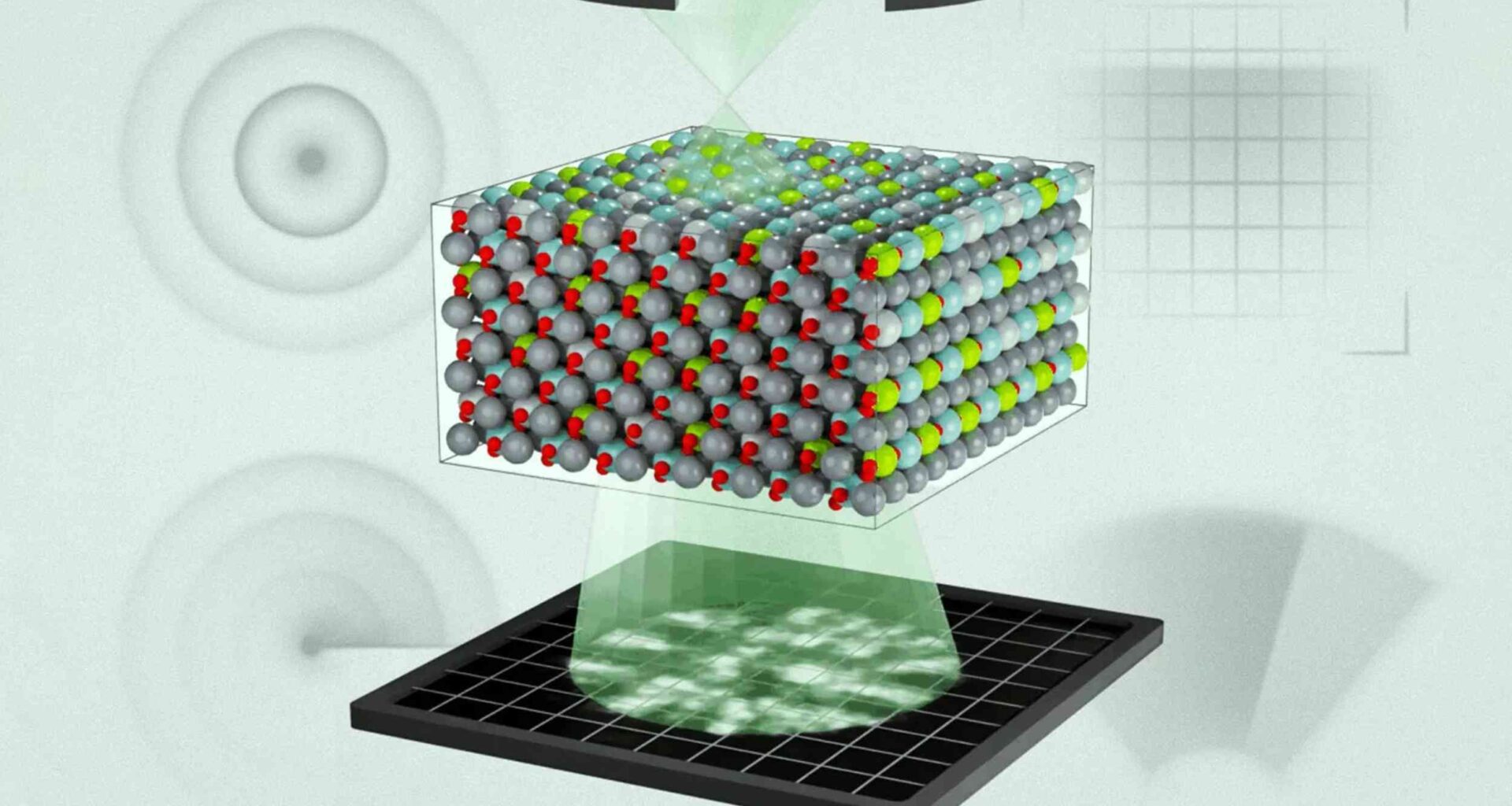
To finally see inside a relaxor, LeBeau’s team turned to electron ptychography. The method works by moving a tightly focused beam of electrons across the crystal’s surface. As the beam moves, sensors below capture the pattern of scattered electrons at each position.
The trick is in the math. Each scan position overlaps with its neighbors, and an algorithm uses the overlap to reconstruct what the electrons passed through, slice by slice.
What pops out is a three-dimensional map of every atom – including which element occupies each position. The method had been demonstrated on thinner materials before. Stretching it to a bulk relaxor was the harder problem.
Examining PMN-PT
When the team applied the technique to a film of PMN-PT, the polar slush stopped being theoretical. The 3D map revealed a continuous patchwork where the direction of electric charge shifted gradually from one region to the next across the crystal.
The regions were smaller than every previous simulation had assumed. Much smaller. And they were correlated – neighboring patches were not random, but bent into one another along soft, gradual boundaries.
“Previously, these models basically had random regions of polarization, but they didn’t tell you how those regions correlate with each other,” said Michael Xu, an MIT postdoc and co-first author.
Chemistry drives polarization
What really surprised the team wasn’t how the slush looked. The chemistry behind it. PMN-PT contains a mix of metal ions: lead, magnesium, niobium, and titanium.
Magnesium has a 2+ charge, niobium a 5+. Those numbers normally feel like dry chemistry. In the reconstruction, they turned out to be the steering wheel.
Where magnesium atoms sat, the surrounding electric charge consistently tilted toward them. Niobium did the opposite – the charge around those ions tilted away. Magnesium was pulling, niobium was pushing.
The random scatter of these ions across the crystal – chemical disorder, in materials-science terms – was setting the entire polar landscape. Earlier models had treated that disorder as background noise.
Better models, better devices
Once the team put those chemistry findings into their computer simulations, the gap between theory and experiment closed sharply. The new model accounts for how specific atoms steer polarization, instead of assuming a random arrangement.
That changes the design game for anyone building ultrasound imaging probes, energy storage capacitors, or solid-state memory. The disorder a designer tolerates in the crystal now has predictable consequences for the device.
“Now that we have a better understanding of exactly what’s going on, we can better predict and engineer the properties we want materials to achieve,” said LeBeau.
PMN-PT and polar slush
Until this study, the polar slush was a hypothesis with strong indirect support. It is now a structure researchers have directly seen in three dimensions inside a working relaxor.
The slush’s geometry, domain sizes, and the chemistry behind its arrangement have all been mapped at the atomic scale for the first time. That gives engineers a target.
Tuning a relaxor’s chemistry to favor specific polarization patterns is no longer a blind guess against an averaged signal – it is an adjustment against a 3D picture.
Ultrasound transducers, sonar projectors, and high-density actuators can now be designed against the structure that actually exists, not the one theorists hoped was there.
The study is published in Science.
—–
Like what you read? Subscribe to our newsletter for engaging articles, exclusive content, and the latest updates.
Check us out on EarthSnap, a free app brought to you by Eric Ralls and Earth.com.
—–