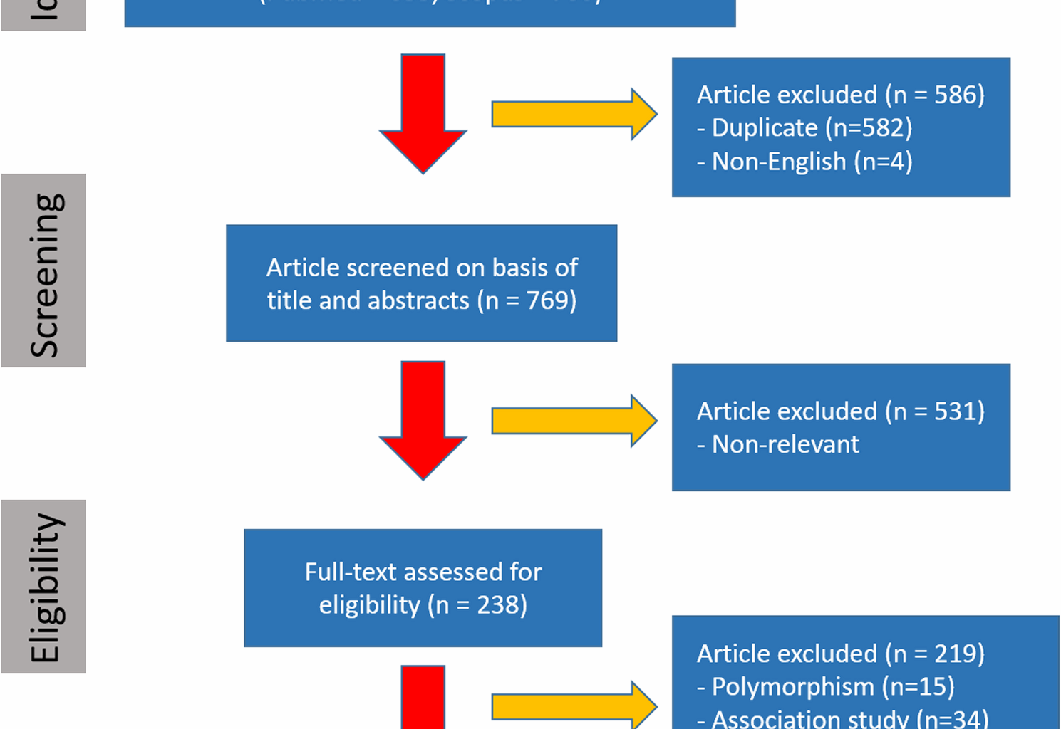
The AXIN2 variants are a common genetic cause of tooth agenesis and may also be associated with an increased risk of cancer. Our systematic review analyzed 34 variants in 91 patients with AXIN2 variants, revealing new insights into AXIN2 genotype-phenotype correlations.
AXIN2 variants are enriched within the SMAD3 domain, suggesting this region is a potential mutational hotspot. Specifically, 20 of 34 identified AXIN2 variants (59%) and 61 of 91 patients (67%) with AXIN2 variants harbor alterations within this domain. The AXIN2 SMAD3 domain interacts with SMAD3, a transcription factor activated by TGF-β signaling. This pathway regulates fundamental cellular processes including proliferation, differentiation, and apoptosis [36,37,38].
SMAD3 plays a crucial role in dento-craniofacial development [39]. Disruption of SMAD3-mediated signaling can affect tooth patterning and morphogenesis, potentially resulting in tooth agenesis [39]. For instance, variants in the SMAD3 gene itself cause Loeys-Dietz syndrome, which can manifest as congenitally missing teeth [40]. Perturbation of the AXIN2 SMAD3 domain could partially disrupt these developmental processes.
In cancer, SMAD3’s role is complex. While canonical TGF-β/SMAD3 signaling acts as a tumor suppressor by inhibiting proliferation and promoting apoptosis in early stages [41, 42], dysregulation of this pathway in advanced cancers can promote invasion, migration, and metastasis in cancers such as colorectal, breast, and lung cancer [41, 42]. Therefore, alterations within the AXIN2 SMAD3 domain could potentially contribute to both tooth agenesis and cancer predisposition through disruption of SMAD3 function. Further research is needed to elucidate the precise mechanisms by which these AXIN2 variants affect SMAD3 signaling and contribute to these phenotypes.
Interestingly, the observation that 86.4% of patients with AXIN2 variants in the SMAD3 domain exhibited tooth agenesis, while patients with variants in the β-catenin domain did not, presents an intriguing paradox. β-catenin is a key mediator of Wnt signaling, a pathway critically involved in tooth development [43, 44]. The Wnt/β-catenin signaling pathway is dynamically active throughout all stages of tooth development, from placode formation to the bell stage [43, 44]. Therefore, the disruption of AXIN2’s interaction with β-catenin might be expected to affect tooth development. Several hypotheses could explain this finding. First, the AXIN2 variants in the β-catenin binding domain may not significantly impair β-catenin binding or Wnt signaling, requiring functional studies for confirmation. Second, compensatory mechanisms within the Wnt pathway or other signaling pathways may mitigate the effects of these variants. Third, the distinct roles of SMAD3 and β-catenin in tooth development could mean that disruption of SMAD3 signaling has a greater impact on tooth agenesis. Finally, the small sample size of patients with β-catenin domain variants (4 patients and only 1 variant) may limit statistical detection of tooth agenesis. Larger studies are needed to confirm these findings and explore the molecular mechanisms involved.
When categorized by variant type, patients with nonsense variants exhibited the highest average number of missing teeth. This observation correlates with the complete penetrance of tooth agenesis observed in this group; all 16 individuals with nonsense variants exhibited missing teeth. In contrast, incomplete penetrance was observed in patients with missense or frameshift variants, some of whom did not exhibit tooth agenesis. Nonsense variants typically result in premature truncation of the AXIN2 protein, leading to a complete loss of function. This likely explains the higher penetrance and more severe phenotype observed in these patients compared to those with missense or frameshift variants, which may retain some residual protein function. Further investigation into the specific effects of different variant types on AXIN2 protein function and downstream signaling pathways is warranted to fully elucidate the genotype-phenotype correlation. Tooth agenesis exhibited a largely symmetrical pattern between the maxilla and mandible, with comparable prevalence on the left and right sides, except for the first premolars and incisors. Second premolars showed the highest prevalence of agenesis, while maxillary central incisors were least affected. These findings are consistent with previous reports on AXIN2-related tooth agenesis, which also identified second premolars as the most frequently missing tooth type (61.5–74.4%) and maxillary central incisors as the least (7.7%) [45]. A high rate of third molar agenesis (80.8–84.6%) was observed in a subset of 26 patients with detailed records. Details are shown in Supplementary Table S3. However, these data were excluded from the overall agenesis pattern analysis as the absence of third molars is often considered a normal variation or an evolutionary adaptation rather than a developmental anomaly [46, 47].
The pattern of tooth agenesis associated with AXIN2 variant carriers differs from that associated with other genes implicated in tooth development, such as PAX9 and MSX1. Individuals with PAX9 variants, second molars are most commonly affected (78% agenesis), while mandibular first premolars are least affected (4%) [17]. Conversely, MSX1 variants are predominantly associated with second premolars agenesis (83–86%), with canines being the least affected (6–8%) [45, 48]. This suggests distinct roles for AXIN2, PAX9, and MSX1 in regulating tooth development, potentially influencing the development of specific tooth types.
The consistency of the dental phenotype data across the cohort supports the reliability of our analysis. As shown in the Results, the average number of missing teeth in the subgroup with complete records was nearly identical to that of the overall cohort. This strong concordance suggests that the subgroup used for detailed pattern analysis (e.g., Fig. 4) is representative of the broader population, thereby reinforcing the robustness of our conclusions regarding the AXIN2-associated dental phenotype.
However, because a significant portion of patients had incomplete dental or cancer data, the generalizability of our findings remains limited. Missing cancer testing records for 27 patients may bias incidence estimates toward individuals with greater medical follow-up, while incomplete dental charts could obscure subtle variations in tooth agenesis patterns. These limitations underscore that our results should be interpreted with caution and highlight the need for future studies with comprehensive, systematic dental and oncologic assessments.
Regarding cleft lip or palate, only one article in our review reported three patients with cleft palate from the same family [35]. One of these patients had two missing teeth, but none had cancer, potentially due to young age. Previous studies, such as those by Mostowska, et al.. (2012), found borderline association between AXIN2 variants and a decreasing risk of cleft lip or palate [49], although Motahari’s research found a higher frequency of cleft lip or palate in Asian patients with the G allele and AG or AG + GG genotypes of AXIN2 [50]. More studies are needed to clarify this potential association.
An intriguing aspect of AXIN2-related syndrome is the apparent paradox wherein Wnt signaling hyperactivation, which drives tumorigenesis, also leads to tooth agenesis [8]. This is particularly notable given the reported role of Wnt pathway activation in causing supernumerary teeth [44, 51]. The fact that variants leading to a hyperactive Wnt signal can result in both missing and extra teeth highlights the critical importance of the signal’s magnitude and precise spatiotemporal regulation during development. While a controlled increase in Wnt signaling may be sufficient to induce additional tooth structures, the loss of a key negative regulator like AXIN2 is hypothesized to cause a pathological over-activation [8]. This excessive, unregulated signal likely disrupts the finely tuned cues required for the initial formation and progression of the tooth germ. Therefore, the developmental outcome of Wnt pathway dysregulation is not a simple dose-response but depends on whether the signal provides a regulated instruction or a complete disruption of the developmental program. This dual role exemplifies the powerful yet delicate nature of Wnt signaling in both organogenesis and disease.
Besides tooth agenesis, AXIN2 variants have been linked to an increased risk of cancer, as Wnt signaling pathway is essential for cellular homeostasis and proliferation regulation [14]. Variants in AXIN2 can lead to aberrant Wnt signaling activation, promoting uncontrolled cell growth and tumorigenesis. Our analysis showed a high prevalence of colorectal cancer among both male and female AXIN2 variant carriers, consistent with other studies showing a link between tooth agenesis and colorectal cancer [8, 13, 14]. It has also been observed in previous studies that oligodontia and predisposition to cancer are caused by a mutation of Arg656*, in the Wnt-signaling regulator AXIN2. Also in a separate case of severe tooth agenesis a de novo frameshift mutation 1994-1995insG in AXIN2 was observed [8]. Both the above-mentioned variants have been found to be responsible for the activation of Wnt signaling.
Our study identified c.1966 C >T (p.Arg656*) and c.1994dupG (p.Asn666Glnfs41) as the two most common AXIN2 variants. Both variants introduce premature stop codons, which are predicted to result in a truncated protein that lacks the critical SMAD3 and DIX domains [8]. The pathogenicity of these specific variants is well-established in the literature. For example, c.1966 C >T has been shown to co-segregate with both oligodontia and colorectal cancer predisposition in a Finnish family [8], and both variants are classified as pathogenic in ClinVar. This is mechanistically supported by prior work indicating that truncating variants in exon 8, including p.Arg656*, lead to elevated β-catenin/TCF signaling, consistent with a loss of AXIN2 scaffold function [24]. Together, these data from our cohort and the literature provide strong evidence that these variants exert their functional impact through the disruption of Wnt signaling, thereby offering a plausible molecular basis for the co-occurrence of both dental and oncologic phenotypes.
Although genome-wide studies by Williams, et al. (2018) suggested several genes associated with colorectal cancer may also influence tooth development (e.g., ATF1, DUSP10, and CASC8), AXIN2 was not among the genes they identified [52]. This may reflect limitations in the power of GWAS to detect genes with moderate effects or those involved in rare syndromes. In the present study, female AXIN2 variant carriers also presented with breast and ovarian cancer, consistent with previous studies [53, 54]. Additionally, skin cancer was observed in both males and females carriers [55]. In another report a family was observed to have presented with oligodontia and colorectal adenomatous polyposis. Three members of the family were reported to have c.1972delA, p.Ser658Alafs*31 nonsense variant in AXIN2. The three members had a mean of 16.5 teeth and a mean of 49 polyps which also highlighted the need to confirm phenotypic information in cases with familial polyposis [25]. This further underscores the pleiotropic nature of AXIN2 and its potential involvement in diverse developmental and disease processes.
A striking co-occurrence of tooth agenesis and cancer was observed among AXIN2 variant carriers, especially those with variants in the SMAD3 domain. All but one patient presenting with both cancer and tooth agenesis harbored a variant within this domain. The single exception carried a variant located in a currently unclassified region of the AXIN2 gene. This observation strengthens the hypothesis that disruption of the SMAD3-AXIN2 interaction plays a significant role in the pathogenesis of both conditions.
Although age remains a major determinant of cancer risk, the observation of tooth agenesis, particularly when presenting as oligodontia, may serve as an early clinical indicator of an underlying AXIN2 variant. In our review, the average age of colorectal cancer onset in affected individuals was younger than that typically reported in population-based studies [56], suggesting a possible contribution of hereditary factors. The presence of tooth agenesis in such cases may support consideration for genetic evaluation, especially when there is a relevant family history. While these findings are not intended to directly inform clinical protocols, they highlight the need for further research into genotype-phenotype correlations and their potential role in early risk stratification.
A limitation of this study is the potential for ascertainment bias related to the age of onset of different clinical features. As our analysis shows, patients with AXIN2 variants are often identified in childhood or adolescence due to tooth agenesis. The mean age of patients in the cohort included in this review with only a dental phenotype was significantly lower than that of patients with a cancer diagnosis. Consequently, many of these younger patients may not have yet reached the age at which cancer risk becomes substantial. This age-dependent penetrance means that the prevalence of cancer reported in this collected cohort is likely an underestimation of the true lifetime risk. While the available data precluded a formal sensitivity analysis, an illustrative projection based on published age-specific colorectal cancer incidence rates indicates the potential magnitude of this bias. Given that colorectal cancer incidence increases by more than an order of magnitude between the mean age of our dental-only cohort and that of our cancer-diagnosis group, our findings likely represent a substantial lower-bound estimate of lifetime penetrance. Longitudinal follow-up of young patients identified with pathogenic AXIN2 variants is essential to accurately determine the lifetime cancer risk and establish appropriate surveillance guidelines. Furthermore, it is important to note that the data compiled from the selected articles reflects the patient’s age at the time of reporting, which may not correspond to the age of cancer diagnosis, a data point that was not uniformly available in the source literature.

