
Scientists investigating the fundamental mechanisms of morphogenesis have long recognised the importance of cell adhesion in shaping developing tissues. Adrian Aguirre-Tamaral, Elisa Floris, and Bernat Corominas-Murtra, all from the Department of Biology at the University of Graz, demonstrate a crucial link between cell-cell adhesion and the resulting topology of early embryonic tissues. Their research reveals that a localised property at cell contacts exerts a significant influence on the global geometry and characteristics of developing embryos. This finding is particularly significant as it establishes how local cellular interactions can drive large-scale tissue organisation, providing new insight into the physical forces underpinning organ formation.
This work demonstrates that the arrangement of cells and their connections, the tissue’s topology, plays a critical role in defining both the geometry and material characteristics of developing embryos.
Researchers established that changes in cell adhesion directly influence the topology of cell networks, ultimately shaping the morphogenesis of tissues and organs. The study identifies a dynamic interplay between adhesion and topology as a primary driver of embryonic development, offering new insights into the physical principles governing organ formation.
The research focuses on understanding how tissues acquire complex shapes without external forces, highlighting the importance of coordinated cellular behaviour and changes in mechanical properties. Investigations reveal that topological properties, such as the presence of holes or tubular structures, are fundamental characteristics of embryonic tissues, acting as constraints on potential geometries.
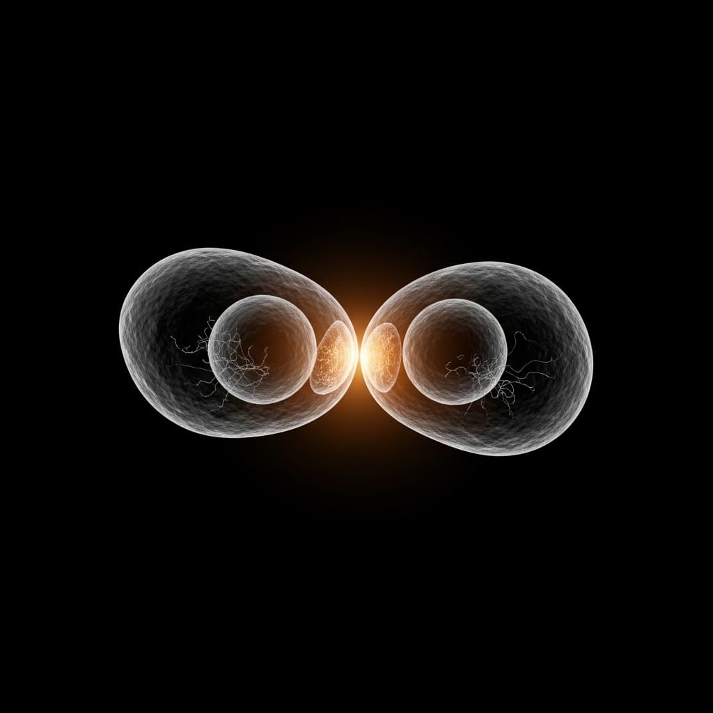
By examining the network of cell-cell contacts, scientists defined tissue topology as a network structure based solely on cell connections, irrespective of geometric details. Different arrangements of cells create distinct topological configurations, each described by an adjacency matrix representing contact relationships.
Specifically, the study demonstrates that topological transitions, represented by changes in this adjacency matrix, are directly linked to alterations in cell adhesion. Cell adhesion, far from being a simple glue, is presented as a highly regulated mechanochemical process coordinating tissue structure and maintenance.
Researchers utilised a simplified model of cell membranes, drawing parallels with foam physics, to analyse the interplay between cell-cell and cell-fluid surface tensions. A non-dimensional parameter, α, defined as the ratio of cell-cell to cell-fluid surface tension (γcc/(2γcf)), was identified as a key variable governing this relationship.
This parameter, α, can be empirically determined from the contact angle between cells and fluid, following the Young-Dupré relation, providing a testable link between theory and experiment. A Hamiltonian function was developed to describe the energy of a cell configuration based on adhesion, cell area in contact with fluid, and cell compressibility, further elucidating the relationship between topology, adhesion, and tissue mechanics. The findings suggest that this interplay between cell adhesion and topology is a central mechanism driving morphogenesis and establishing functional phenotypic traits.
Computational modelling of cell adhesion using Surface Evolver and principles of sphere packing
A detailed analysis of cell-cell adhesion was undertaken to investigate its role in embryonic tissue morphogenesis. Researchers employed computational modelling, specifically utilising the Surface Evolver software, to simulate cell arrangements and adhesion properties. This program allowed for the minimisation of energy within a system of cells, effectively modelling how cells pack and interact based on their adhesive forces.
Simulations were performed on cell clusters to determine the impact of varying adhesive strengths on overall tissue geometry and mechanical properties. The study drew inspiration from established principles of sphere packing, referencing work on minimal energy clusters of hard spheres with short-range attractions to inform the modelling of cell arrangements.
Specifically, the research considered concepts from disordered 2D froths and random packings of frictionless particles to understand how cells behave when subjected to external forces and internal stresses. By manipulating the adhesive properties within the simulations, researchers observed transitions between different states of tissue organisation, mirroring the rigidity transitions seen in developing embryos.
Furthermore, the work connected these computational findings to observations in developing zebrafish embryos, utilising established staging criteria to correlate simulated tissue behaviours with actual embryonic development. This allowed for validation of the model and provided insights into how changes in cell adhesion contribute to the formation of complex organ geometries. The research also considered the influence of 3D matrix confinement on cell behaviour, building on previous studies demonstrating jamming transitions in breast cancer invasion.
Adhesion and density define distinct embryonic tissue architectures through stress propagation
Researchers demonstrate that a localised, pairwise property at cell-cell contacts significantly influences embryonic tissue geometry and characteristics. Numerical experiments using Surface evolver software reveal that stress propagation, measured as the relative increase of internal pressure in cells, is substantially larger in tissues below an adhesion critical point, αc, compared to those above it.
Specifically, tissues with cell fractions exceeding a jamming critical point, φc, exhibit markedly different stress propagation scales depending on whether they are above or below αc. The study establishes a phase diagram defined by adhesion, α, and density, φ, comprising four distinct regions surrounding a double critical point at (αc, φc).
These regions correlate with different tissue architectures: epithelial-like when α φc, lumen-like when α αc and φ φc, and sparse mesenchymal-like when α αc and φ The research highlights that tissues with the same a-priori cell fraction, φ, greater than 0.84, display differing stress responses based on their adhesion state. Tissues below αc exhibit a broader scale of stress propagation, indicating a less rigid structure, while those above αc demonstrate a more constrained response.
This work provides abstract tools to connect microscopic cell-cell interactions with macroscopic tissue structure and properties, offering insights into how developing organisms regulate tissue topology and phenotypic outcomes. Further investigation will focus on linking these topological and mechanical properties to cell fate decisions and pathological scenarios, bridging function and morphogenesis.
Adhesion and density govern mechanical behaviour in modelled embryonic tissues
Cell adhesion plays a crucial role in orchestrating the complex physical changes necessary for proper organ and tissue formation during embryonic development. Investigations into this process reveal that localised interactions between cells have significant, far-reaching consequences for the overall geometry and material properties of developing tissues.
Specifically, the research demonstrates that the arrangement of cells and their adhesive forces define a tissue’s structural characteristics and how it responds to physical stress. Numerical modelling using computational tools indicates that tissues with similar cell densities exhibit differing stress propagation depending on the degree of cell adhesion.
Tissues with stronger adhesion display limited stress transmission, while those with weaker adhesion show a broader scale of internal pressure changes when compressed. This relationship is encapsulated in a phase diagram defined by adhesion strength and cell density, which identifies four distinct tissue architectures, epithelial-like, lumen-like, and two mesenchymal-like states, each with unique structural and material properties.
The theoretical framework presented connects microscopic cell-cell interactions to macroscopic tissue behaviour, offering a means to understand how tissue topology influences development. While this work concentrates on cell adhesion, the authors acknowledge that further research is needed to link these topological and mechanical properties to cell fate decisions and disease processes. Future studies should explore the interplay between tissue mechanics, cell function, and morphogenesis to gain a more complete understanding of developmental biology.