
Scientists have created a three-dimensional “heart-on-a-chip” (HOC) that could provide a breakthrough in the fight against the world’s leading cause of death, cardiovascular disease.
One major challenge is that we cannot easily test how a human heart will react to a drug or disease without putting someone at risk. This engineered heart tissue beats on its own, it mobilizes calcium to initiate muscular activity, and it responds predictably to common drugs.
It’s the first to incorporate a dual-sensing platform that provides real-time tracking of activity throughout the heart tissue down to the cellular level.
In a recent paper, scientists from multiple Canadian institutions describe how they achieved this “significant advance in cardiac tissue engineering and pharmacological testing.”
The key advance here is the integration of sensors that can detect both macro-scale and micro-scale cardiac activity. Both current HOC platforms and the research team’s previous iteration, described in a 2024 paper, lack high-resolution cellular-level sensing.
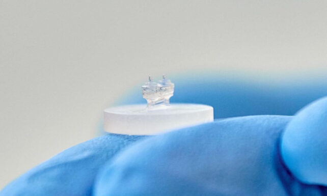 In previous research, the researchers created “hearts on a chip,” ring-shaped devices 3D-printed using a bio-ink containing the patient’s own stem cells. (Véronique Lavoie, CHU Sainte-Justine)
Small-scale sensing is vital because many cardiovascular diseases (CVDs) are associated with dysfunction in cardiomyocytes, the individual contractile cells that form heart muscle tissue, or myocardium. As a result, measuring cellular function is critical for preventing heart failure in patients with CVDs.
To build their HOCs, the researchers harvested cardiac muscle cells and cardiac connective tissue cells from rats. They inserted these cells into a gel-like matrix rich in fibrous proteins and nutrients to stimulate growth, and then seeded them on tiny, flexible silicon-based chips.
The researchers embedded two types of sensors in their HOCs. To measure macro-scale forces, they sandwiched the engineered heart tissues between two elastic pillars. These pillars deform with each heartbeat, and the amount of deformation corresponds to the contractile strength throughout the tissue.
The researchers also immersed flexible, hydrogel-based microsensors within the tissue. The deformation of these tiny droplets, averaging just 50 micrometers (0.002 inches) in size, captures local mechanical stresses at the cellular level.
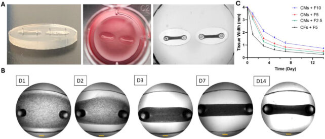 The dense heart tissues formed in vitro are suspended between two flexible, elastic pillars that measure the contractile force of the engineered cardiac muscle. (Mousavi et al., Nano Micro Small, 2025)
This is a major step toward testing pathologies in vitro, since cell-generated forces govern the fate of cardiac tissues, including their formation, remodeling, contractile efficiency, wound healing, and cancer progression.
In vitro testing can also inform pharmacological interventions. So the researchers assessed the feasibility of using their HOCs to perform drug screening, by treating them with two well-studied compounds.
The first, norepinephrine, is also known as noradrenaline. It primes the body’s fight-or-flight response and is used in healthcare settings to increase heart activity and maintain blood pressure, including during cardiac arrest.
To test the opposite effect and decrease contractile activity, the researchers also applied blebbistatin, an inhibitor of muscle activity.
The drugs worked as predicted, demonstrating that the HOCs can forecast how cardiac force generation and heart rhythms respond to common compounds.

“The ability to observe the tissue’s response to different compounds in real time represents a major advantage for preclinical development and translational research,” says first author Ali Mousavi, a biomedical engineer at the University of Montreal.
Next, the researchers plan to simulate specific disorders by building heart tissues using cells from patients living with various cardiac conditions. These include dilated cardiomyopathy, an often-genetic heart muscle disease that can lead to heart failure, as well as arrhythmias, an umbrella of disorders that cause heart rate abnormalities.
Related: This Beating Sesame Seed-Sized ‘Human Heart’ Grew Itself in a Lab
In the long run, HOCs could help doctors choose treatments using tests run on a patient’s own cells, before a medication is prescribed.
“This breakthrough brings us even closer to true precision health,” concludes senior author Houman Savoji, a mechanical and biomedical engineer at the University of Montreal, “by giving us the ability to identify the most effective medication for each person before treatment is even administered.”
This research was published in the journal Nano Micro Small.