
Researchers have identified a new class of fluorescent dyes that enable high-resolution cancer imaging in living cells – achieving detail once limited to fixed samples.
The technology reveals how cancer-related processes unfold in real time, exposing molecular behavior that was previously hidden during live imaging.
Fluorescent dyes for cancer imaging

At the edge of a moving cell, tiny grip structures come into view as they form and release during motion.
Dr. Catherine Galbraith, associate professor of biomedical engineering at OHSU, connected specific dye behaviors to the conditions that make these structures visible inside living cells.
The observations showed that the same cellular features can appear differently depending on local crowding, acidity, and motion within the cell.
That variability makes it necessary to match each dye carefully to the biological context before extending the approach to broader cellular systems.
Why the new dyes are better
Ordinary super-resolution microscopy, imaging that beats light’s usual blur, often struggles in living cells for a simple reason.
Many systems depend on intense light or added chemicals to force dyes on and off, and those conditions can stress cells.
That stress matters when researchers need minutes of behavior instead of a single frozen frame, especially in cancer biology.
By blinking naturally, the new dyes removed much of that setup burden and opened advanced imaging to more ordinary labs.
No single winner
Chemical tweaks pushed on-off ratios across two orders of magnitude, giving researchers a panel instead of a single favorite.
Some versions worked better when molecules were packed tightly together, while others handled sparser targets or quicker image sequences.
In fixed cells, lower on-off ratios generally produced cleaner molecular pinpoints, but live cells changed the balance by altering glow timing.
That is why the study argues against a one-size-fits-all label and toward a set chosen by environment.
Watching the spread of cancer
For cancer researchers, the most striking view may be focal adhesions, protein-rich grip points that help cells crawl through tissue.
Those structures form and break apart as cells pull themselves forward, a process tied to invasion and the spread of cancer.
Because metastatic tumor cells must migrate and invade surrounding tissue, the National Cancer Institute (NCI) treats cell movement as a core part of cancer spread.
“These tools let us see critical cancer-related processes happening live, at the very small scale where key decisions are made inside cells,” said Galbraith.
Following gene activity
Inside the nucleus, the dyes also tracked chromatin, DNA wrapped around proteins, while cells remained alive and changing.
That mattered because gene activity depends partly on how tightly this material is packed and whether crowded regions open.
One version also produced sixfold more usable molecular detections than an older light-activated label while tracking how open DNA stayed.
Those gains made it easier to follow gene-control activity without freezing cells at a single moment.
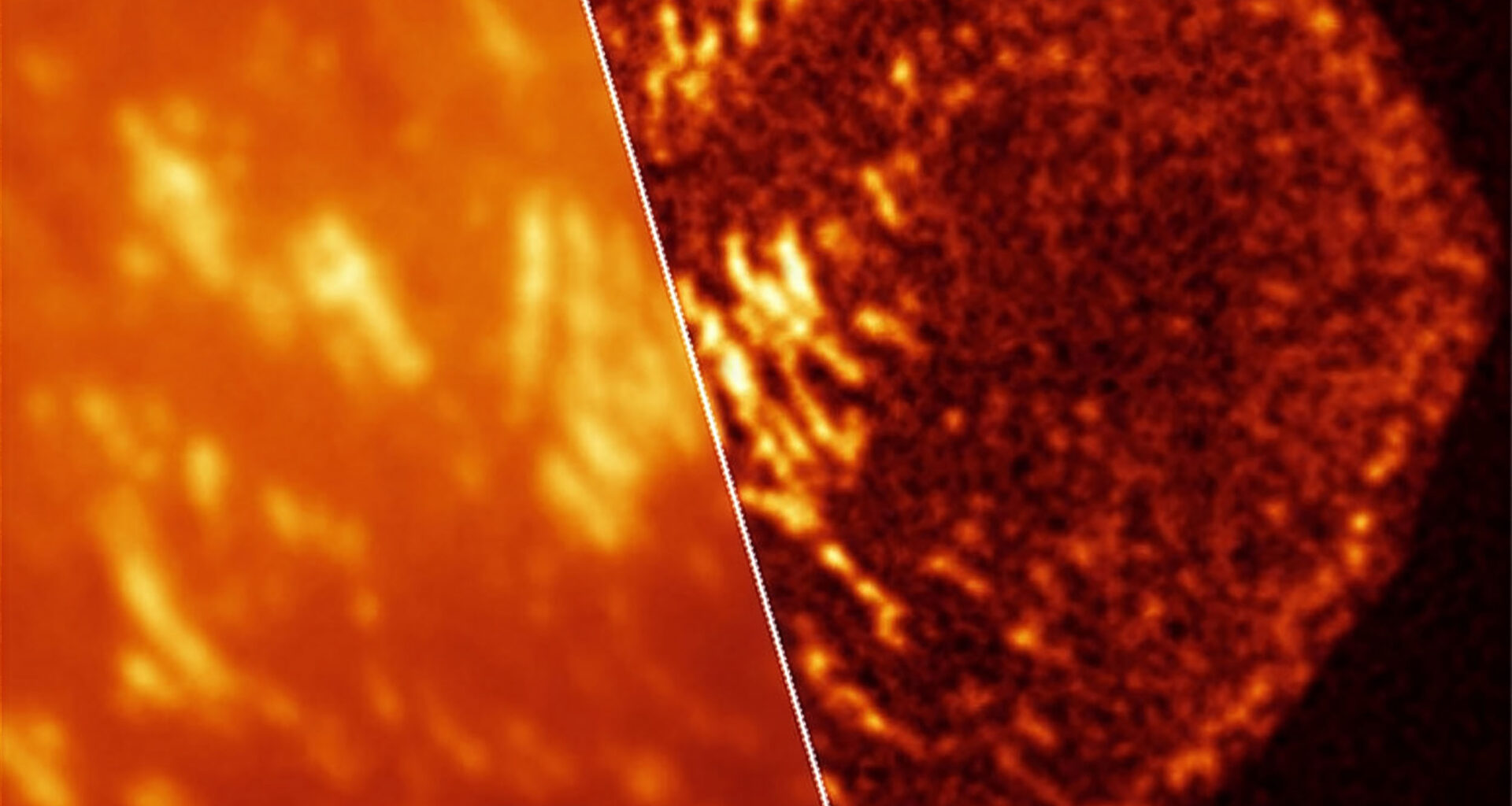
 When cells invade, they grip — and now we can see exactly how. The combination of super-resolution imaging and newly developed spontaneously blinking Janelia Fluor dyes reveal the fine molecular architecture of focal adhesions that live cells use to migrate and invade tissue (right) — detail completely invisible to conventional imaging (left). Credit: Cathy Galbraith/OHSU. Click image to enlarge.Dyes are sensitive to acidity
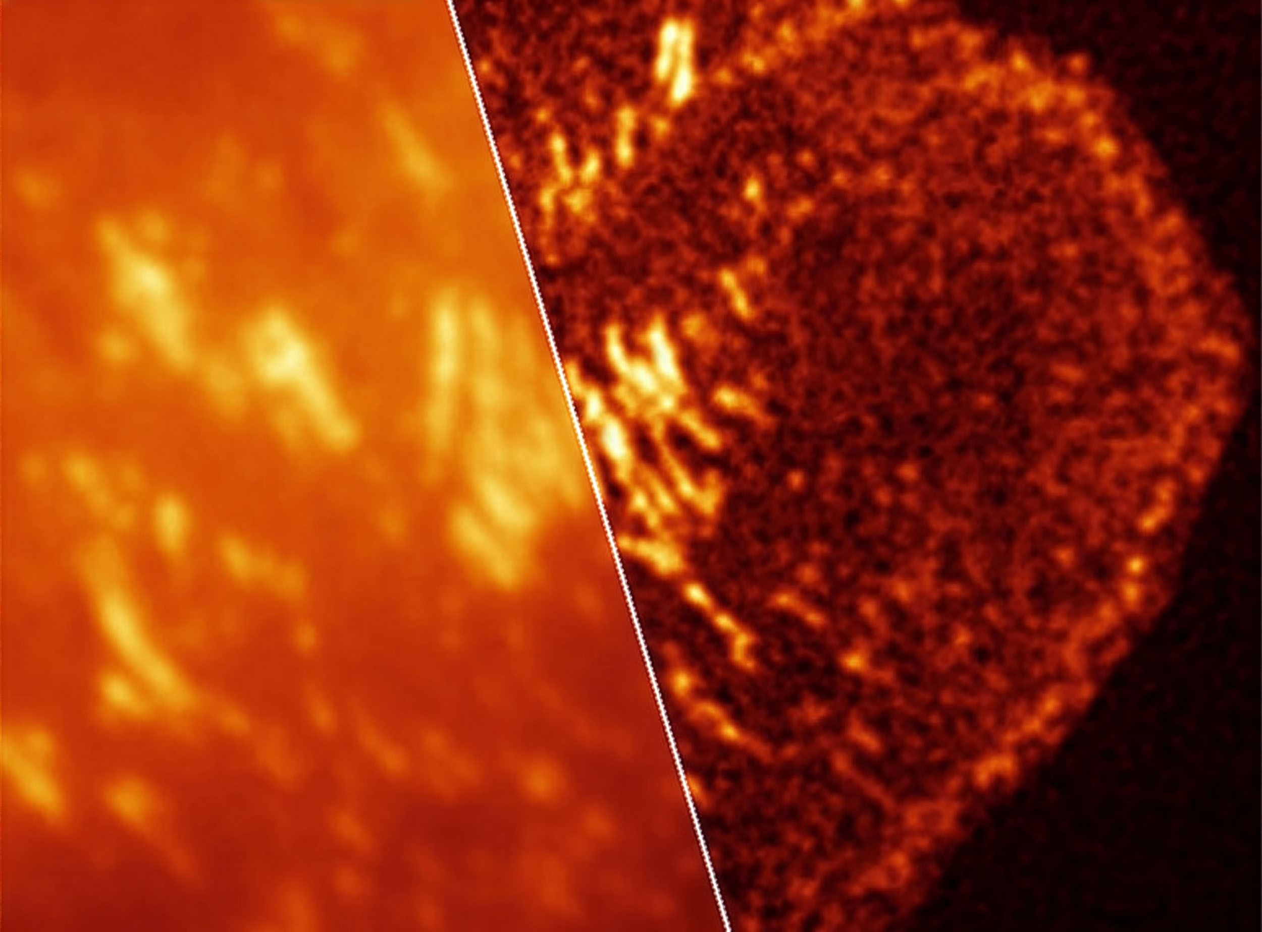
Acidity turned out to matter so much that a dye suited for one compartment could fail in another.
In lysosomes, acidic recycling compartments inside cells, one low-glow dye sharpened images precisely because acid changed its blinking behavior.
Fixing the cells reversed that advantage by neutralizing the compartment, and a different dye then performed better.
Tumors often contain acidic pockets, so this environmental sensitivity could become useful rather than troublesome in cancer studies.
Tuning into RNA messages
Messenger RNA – genetic instructions that carry DNA’s code to build proteins – also appeared as clearer signals when the team paired the blinking dyes with RNA labeling probes.
That let the microscope separate nearby RNA messages more cleanly, which helps scientists tell whether genes act alone or in clusters.
A second imaging mode built sharper pictures from brightness flickers using fewer than 1,000 frames, making quick readouts possible.
For labs tracking RNA while cancer cells turn genes on and off, that speed can widen what fits into routine experiments.
The role of speed and resolution
Speed mattered just as much as sharpness when the researchers tracked moving mitochondria and growing cell attachments over time.
One approach followed individual glowing molecules for the finest detail, while another traded some detail for faster updates.
Near the front edge, the flicker-based method visualized small new attachments, and one live-cell setup ran threefold faster than earlier work.
The results suggest that researchers do not need one perfect technique if they can match resolution and timing to the biology.
Adopting the dyes for cancer imaging
The ease of use may decide whether these dyes matter, because many cancer labs lack custom hardware or patience for delicate image tuning.
“New tools open up biology,” Galbraith said. Even so, the panel is not plug-and-play, since the wrong dye can underperform when density, acidity, or fixation change.
The OHSU team has essentially handed researchers practical instructions, and that may speed adoption.
From moving cell edges to packed DNA and acidic vesicles, the new dyes made live-cell microscopy more flexible.
The study is published in the journal Nature Methods.
—–
Like what you read? Subscribe to our newsletter for engaging articles, exclusive content, and the latest updates.
Check us out on EarthSnap, a free app brought to you by Eric Ralls and Earth.com.
—–