
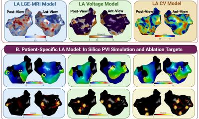
The findings point clearly toward combining all three data types within a single hybrid model as the most promising path forward and lay the scientific foundation for doing so.
Lead author Dr Mahmoud Ehnesh, Postdoctoral Research Assistant at Queen Mary University of London, said: “We found that MRI scans of the heart are valuable, but they don’t tell the whole story. This study compared three types of clinical data, imaging and two types of electroanatomic mapping data, and found that each captures a different dimension of how Atrial Fibrillation behaves in an individual heart. Relying on any single source means missing part of the picture. Combining all three within a single personalised model is the most promising path toward more accurate, targeted ablation for persistent AF patients.”
Senior author Dr Caroline Roney, Reader in Computational Medicine at Queen Mary University of London, said: “If you have a persistent irregular heartbeat (Atrial Fibrillation) and are considering ablation, personalised computer modelling may one day help surgeons plan your procedure more precisely, but this technology is still in the research phase and not yet part of routine clinical care.”
The team working on Comparative Multimodal Calibration of Patient-Specific Atrial Fibrillation models included researchers at Queen Mary University of London, Royal Brompton & Harefield Hospital (Guy’s and St Thomas’ NHS Foundation Trust), Imperial College London, King’s College London, the University of Leeds, and IHU Liryc (Bordeaux).
Source: Queen Mary University of London