
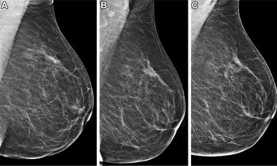
The increase in scores among cancer patients was detectable as early as six years prior to diagnosis and became more pronounced over time
Constance D. Lehman
A validated, open-source image-only deep learning model was applied to all the mammograms to generate continuous five-year breast cancer risk scores. No demographic, clinical or historical imaging data were used.
A total of 817 women (1%) were diagnosed with breast cancer within 365 days of their index exam, including 451 (55%) with invasive cancer, 118 (14%) with ductal carcinoma in situ (DCIS), and 248 (30%) with an unknown cancer. Of these, 682 (83%) were screen-detected cancers, and 135 (17%) were interval cancers. The remaining 53,197 women (98%) were not diagnosed with breast cancer during follow-up and were categorized as cancer-free controls.
The researchers compared the risk scores of the 817 women diagnosed with invasive cancer or DCIS with the scores of the 53,197 cancer-free control individuals.
“We observed clinically relevant differences in risk trajectories between women who did and did not develop cancer,” Dr. Lehman said. “The increase in scores among cancer patients was detectable as early as six years prior to diagnosis and became more pronounced over time.”
Among the cancer patients, AI risk scores increased progressively over the six years preceding diagnosis, with the median score increasing from 2.1 in the first five to six years of the study period to 6.6 at the index exam. Cancer-free women exhibited stable scores across all time points, with medians ranging from 1.8 to 2.2 over the study period.
“These findings demonstrate signals, invisible to the human eye, in the image alone can predict future risk,” Dr. Lehman said. “This is exciting, because 85% of women diagnosed with breast cancer do not have a significant family history of breast cancer or known genetic mutations.”
Dr. Lehman noted that the majority of breast cancer cases are sporadic, meaning they’re not driven by familial inheritance or genetics. “AI-derived risk scores can identify patients who are otherwise predisposed to the disease, and our findings demonstrate that image-based AI risk scores evolve over time and that changes in those scores may provide additional information about future breast cancer risk,” she said.
The risk score trajectory among cancer patients increased most steeply in the years immediately preceding the diagnosis. A gradual increase in scores in the early years of the study period was followed by a much steeper increase two years prior to the index exam. In contrast, the cancer-free trajectories remained essentially flat across the study period.
“These trends remained robust across subgroups defined by age and breast density, further supporting the generalizability of our findings,” Dr. Lehman said. “This is particularly relevant given persistent disparities in screening performance across patient populations. A dynamic biomarker approach grounded in the imaging data could mitigate some of these disparities by enabling risk-based personalization that does not rely on self-reported or inconsistent clinical data.”
Dr. Lehman said the study’s findings support the potential of image-based risk models as dynamic imaging biomarkers to guide personalized, risk-reduction strategies. “With the power of AI, computer vision, and the ability to extract predictive data, we are able to apply the power of imaging to risk assessment and preventing disease from developing,” she said. “Having a dynamic risk score opens up a whole new domain of more effective preventive therapies for breast cancer, similar to how we screen for and treat patients with high cholesterol and hypertension.”
AI image-based risk scores are incorporated into the 2026 National Comprehensive Cancer Network guidelines. The guidelines recommend that beginning at age 35, women with an elevated five-year risk score (greater than 1.7%) consider breast MRI in addition to annual mammography.
An FDA-approved AI-based, image-based five-year risk-scoring model is currently in clinical use at select healthcare institutions across the U.S.
Source: Radiological Society of North America