
© BASILICOSTUDIO STOCK/stock.adobe.com
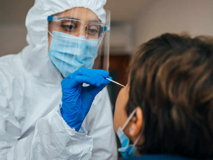
The race to find a COVID-19 vaccine underlined the importance of understanding disease pathways. One promising area of research has been the identification – early and in detail – of changes in cell structure following penetration by a virus.
At the moment, a novel technology capable of capturing this is soft X-ray microscopy (SXM). The microscope works like a CT scanner, but at the cellular level.
“SXM is really cool,” says CoCID(opens in new window) project coordinator Nicola Fletcher from University College Dublin(opens in new window) (UCD) in Ireland. “It fits in between what we call light microscopy – ordinary microscopy – and electron microscopy, which looks in detail at single aspects of a cell. The beauty of SXM is that it takes a whole cell and looks at it in beautiful detail.”
The problem however is that the illumination required for a soft X-ray microscope requires a football-stadium sized facility, called a synchrotron. “There are only six in the world, and these are extremely hard to access,” adds Fletcher.
Miniaturising soft X-ray illumination
The EU-funded CoCID project sought to address this challenge by building on a solution developed by SiriusXT(opens in new window), a spin-off company from UCD. The start-up recently found a way to miniaturise the synchrotron into a small chamber that can fit in a laboratory, providing the same type of soft X-ray illumination as the synchrotron.
The CoCID project sought to further refine the technology and to demonstrate its efficacy in cell structure imaging. The project looked at viruses, which cause structural changes when they infect cells. A consortium including UCD, SiriusXT and various European experts in microscopy and viruses was brought together.
“We had groups studying hepatitis E, SARS-CoV-2, hepatitis C, and herpes virus infections,” explains Fletcher. “Can we understand the structural changes going on when viruses infect cells, and can we reverse these with drugs? These were the questions we wanted to ask.”
Long-term effects of diseases
The CoCID project helped to further refine and develop SiriusXT’s technology, increasing the resolution and bringing the technology closer to the levels of a synchrotron. The project also complemented SiriusXT’s technology with light microscopy.
“Using this technology, we were able to characterise cellular structural changes and identify places that were profoundly changed following infection,” notes Fletcher. “This is a new way of looking at virus-infected cells.”
For example, the project was able to capture images of cells post-viral infection. “We looked at the impact of antiviral therapies,” she says. “Hepatitis C-infected cells look like little junk yards with what we think are parts of dead virus. This opens up new questions such as what this means for the health of cells going forward, and what the long-term effects of diseases like COVID could be.”
Tissue imaging with SXT
SiriusXT’s microscope is now operational at UCD, with several development microscopes available at their own facility in Dublin. “Moving forward, we as researchers want to look more closely at the practical implications of this technology,” adds Fletcher.
This desire has led to a European Research Council(opens in new window) funded project called NanoX, which got underway in June 2025. The idea behind this project is to allow scientists to visualise the complex environment of diseased organs and tissues.
“Our focus in this project is hepatitis E,” explains Fletcher. “We want to try to understand where this virus replicates in the body, which will be critical to designing antiviral treatments in the future.”
Discover other articles in the same domain of application