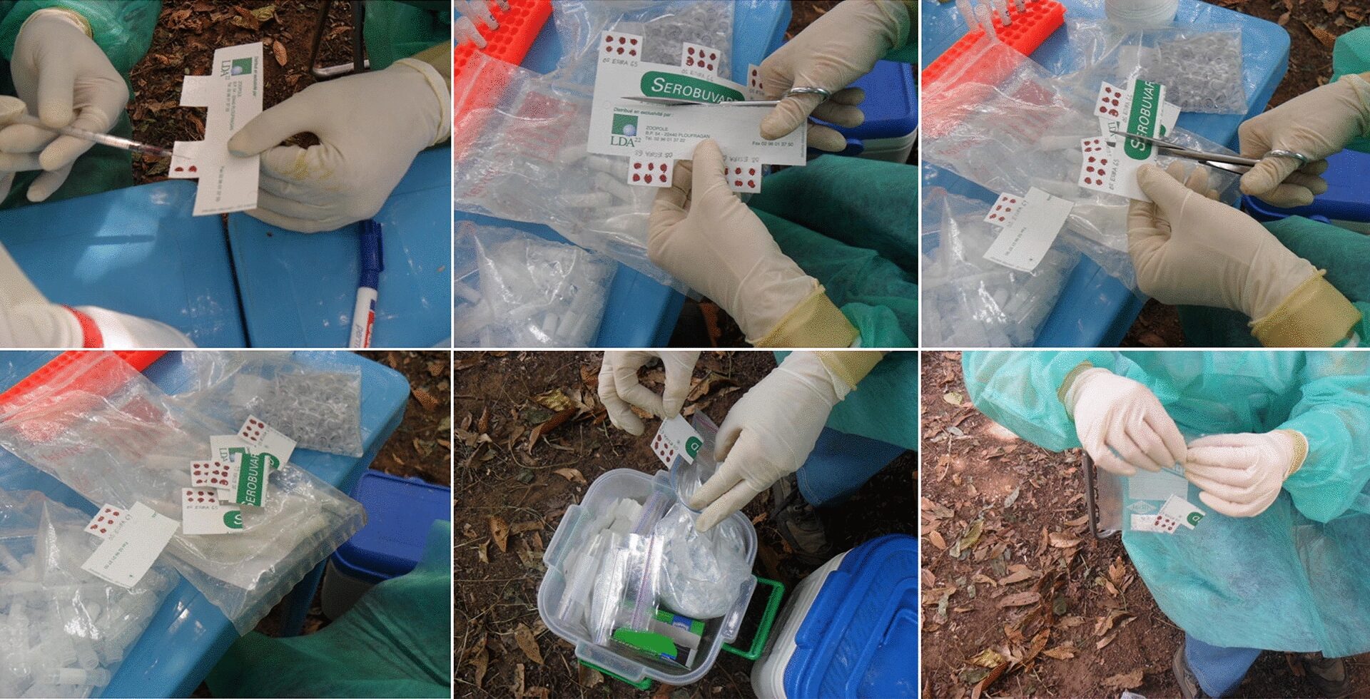
Wang Z, Gerstein MB, Snyder M. RNA-Seq: a revolutionary tool for transcriptomics. Nat Rev Genet. 2009;10:57–63.
Wilhelm BT, Landry JR. RNA-seq-quantitative measurement of expression through massively parallel RNA-sequencing. Methods. 2009;48(3):249–57.
Mortazavi A, Williams BA, McCue K, Schaeffer L, Wold B. Mapping and quantifying mammalian transcriptomes by RNA-seq. Nat Methods. 2008;5(7):621–8.
Li T, Mbala-Kingebeni P, Naccache SN, Thézé J, Bouquet J, Federman S, et al. Metagenomic next-generation sequencing of the 2014 Ebola virus disease outbreak in the Democratic Republic of the Congo. J Clin Microbiol. 2019. https://doi.org/10.1128/JCM.00827-19.
da Costa Castilho M, de Filippis AMB, Machado LC, de Lima Calvanti TYV, Lima MC, Fonseca V, et al. Evidence of Zika virus reinfection by genome diversity and antibody response analysis, Brazil. Emerg Infect Dis. 2024;30(2):310–20.
Yu J, Zhang L, Gao D, Wang J, Li Y, Sun N. Comparison of metagenomic next-generation sequencing and blood culture for diagnosis of bloodstream infections. Front Cell Infect Microbiol. 2024;14:1338861.
Mei JV, Alexander JR, Adam BW, Hannon WH. Use of filter paper for the collection and analysis of human whole blood specimens. J Nutr. 2001;131(5):1631S-1636S.
Lauer E, Widmer C, Versace F, Staub C, Mangin P, Sabatasso S, et al. Body fluid and tissue analysis using filter paper sampling support prior to LC-MS/MS: application to fatal overdose with colchicine. Drug Test Anal. 2013;5(9–10):763–72.
Lehmann S, Delaby C, Vialaret J, Ducos J, Hirtz C. Current and future use of “dried blood spot” analyses in clinical chemistry. Clin Chem Lab Med (CCLM). 2013;51(10):1897–909.
Rahavendran SV, Vekich S, Skor H, Batugo M, Nguyen L, Shetty B, et al. Discovery pharmacokinetic studies in mice using serial microsampling, dried blood spots and microbore LC-MS/MS. Bioanalysis. 2012;4(9):1077–95.
Wickremsinhe ER, Perkins EJ. Using dried blood spot sampling to improve data quality and reduce animal use in mouse pharmacokinetic studies. J Am Assoc Lab Anim Sci. 2015;54(2):139–44.
Henderson CM, Bollinger JG, Becker JO, Wallace JM, Laha TJ, MacCoss MJ, et al. Quantification by nano liquid chromatography parallel reaction monitoring mass spectrometry of human apolipoprotein A-I, apolipoprotein B, and hemoglobin A1c in dried blood spots. Proteomics Clin Appl. 2017;11(78):1600103.
Gao F, McDaniel J, Chen EY, Rockwell HE, Drolet J, Vishnudas VK, et al. Dynamic and temporal assessment of human dried blood spot MS/MSALL shotgun lipidomics analysis. Nutr Metab. 2017;14(1):28.
Demirev PA. Dried blood spots: analysis and applications. Anal Chem. 2013;85(2):779–89.
Zakaria R, Allen KJ, Koplin JJ, Roche P, Greaves RF. Advantages and challenges of dried blood spot analysis by mass spectrometry across the total testing process. EJIFCC. 2016;27(4):288–317.
Guthrie R, Susi A. A simple phenylalanine method for detecting phenylketonuria in large populations of newborn infants. Pediatrics. 1963;32:338–43.
Mei J. Dried blood spot sample collection, storage, and transportation. In: Li W, Lee MS, editors. Dried blood spots. Hoboken: Wiley; 2014. p. 21–31.
Fichet-Calvet E, Rogers DJ. Risk maps of Lassa fever in West Africa. PLoS Negl Trop Dis. 2009;3(3):e388.
Buckley SM, Casals J. Lassa fever, a new virus disease of man from West Africa. 3. Isolation and characterization of the virus. Am J Trop Med Hyg. 1970;19(4):680–91.
Frame JD, Baldwin JM Jr, Gocke DJ, Troup JM. Lassa fever, a new virus disease of man from West Africa. I. Clinical description and pathological findings. Am J Trop Med Hyg. 1970;19(4):670–6.
McCormick JB, King IJ, Webb PA, Johnson KM, O’Sullivan R, Smith ES, et al. A case-control study of the clinical diagnosis and course of Lassa fever. J Infect Dis. 1987;155(3):445–55.
Ogbu O, Ajuluchukwu E, Uneke CJ. Lassa fever in West African sub-region: an overview. J Vector Borne Dis. 2007;44(1):1–11.
Basinski AJ, Fichet-Calvet E, Sjodin AR, Varrelman TJ, Remien CH, Layman NC, Bird BH, Wolking DJ, Monagin C, Ghersi BM et al. Bridging the gap: Using reservoir ecology and human serosurveys to estimate Lassa virus incidence in West Africa. bioRxiv 2020:2020.2003.2005.979658.
Lecompte E, Fichet-Calvet E, Daffis S, Koulemou K, Sylla O, Kourouma F, et al. Mastomys natalensis and Lassa fever, West Africa. Emerg Infect Dis. 2006;12(12):1971–4.
Monath TP, Newhouse VF, Kemp GE, Setzer HW, Cacciapuoti A. Lassa virus isolation from Mastomys natalensis rodents during an epidemic in Sierra Leone. Science. 1974;185:263–5.
Colangelo P, Verheyen E, Leirs H, Tatard C, Denys C, Dobigny G, et al. A mitochondrial phylogeographic scenario for the most widespread African rodent, Mastomys natalensis. Biol J Linn Soc. 2013;108(4):901–16.
Leirs H. Mastomys genus Multimammate mouse. Mammals of Africa: rodents, hares and rabbits, vol. III. 1st ed. London: Bloomsbury Publishing; 2013.
Fichet-Calvet E, Lecompte E, Koivogui L, Soropogui B, Dore A, Kourouma F, et al. Fluctuation of abundance and Lassa virus prevalence in Mastomys natalensis in Guinea, West Africa. Vector Borne Zoonotic Dis. 2007;7(2):119–28.
Stephenson EH, Larson EW, Dominik JW. Effect of environmental factors on aerosol-induced Lassa virus infection. J Med Virol. 1984;14(4):295–303.
Bonwitt J, Saez AM, Lamin J, Ansumana R, Dawson M, Buanie J, et al. At home with Mastomys and Rattus: human-rodent interactions and potential for primary transmission of Lassa virus in domestic spaces. Am J Trop Med Hyg. 2017. https://doi.org/10.4269/ajtmh.16-0675.
ter Meulen J, Lukashevich I, Sidibe K, Inapogui A, Marx M, Dorlemann A, et al. Hunting of peridomestic rodents and consumption of their meat as possible risk factors for rodent-to-human transmission of Lassa virus in the Republic of Guinea. Am J Trop Med Hyg. 1996;55(6):661–6.
Monath TP, Maher M, Casals J, Kissling RE, Cacciapuoti A. Lassa fever in the eastern province of Sierra Leone, 1970–1972. II clinical observations and virological studies on selected hospital cases. Am J Trop Med Hyg. 1974;23:1140–9.
Bonwitt J, Kelly A, Ansumana R, Sahr F, Mari Saez A, Borchert M, et al. Rat-atouille: a mixed method study to characterize rodent hunting and consumption in the context of Lassa fever. EcoHealth. 2016. https://doi.org/10.1007/s10393-016-1098-8.
Olschewski S, Thielebein A, Hoffmann C, Blake O, Muller J, Bockholt S, et al. Validation of inactivation methods for arenaviruses. Viruses. 2021. https://doi.org/10.3390/v13060968.
Magassouba N, Koivogui E, Conde S, Kone M, Koropogui M, Soropogui B, et al. A sporadic and lethal Lassa fever case in Forest Guinea, 2019. Viruses. 2020. https://doi.org/10.3390/v12101062.
Shah R, Barclay ST, Peters ES, Fox R, Gunson R, Bradley-Stewart A, et al. Characterisation of a hepatitis C virus subtype 2a cluster in Scottish PWID with a suboptimal response to Glecaprevir/Pibrentasvir treatment. Viruses. 2022;14(8):1678.
Mills JN, Centers for Disease C, Prevention: Methods for trapping and sampling small mammals for virologic testing. Atlanta: U.S. Dept. of Health & Human Services, Public Health Service, Centers for Disease Control and Prevention; 1995.
Fichet-Calvet E. Chapter 5 – Lassa fever: a rodent-human interaction. In: Johnson N, editor. The role of animals in emerging viral diseases. Boston: Academic Press; 2014. p. 89–123.
Olschlager S, Lelke M, Emmerich P, Panning M, Drosten C, Hass M, et al. Improved detection of Lassa virus by reverse transcription-PCR targeting the 5’ region of S RNA. J Clin Microbiol. 2010;48(6):2009–13.
Vieth S, Drosten C, Lenz O, Vincent M, Omilabu S, Hass M, et al. RT-PCR assay for detection of Lassa virus and related Old World arenaviruses targeting the L gene. Trans R Soc Trop Med Hyg. 2007;101(12):1253–64.
Thomson E, Ip CL, Badhan A, Christiansen MT, Adamson W, Ansari MA, et al. Comparison of next-generation sequencing technologies for comprehensive assessment of full-length hepatitis C viral genomes. J Clin Microbiol. 2016;54(10):2470–84.
Davis C, Mgomella GS, da Silva Filipe A, Frost EH, Giroux G, Hughes J, et al. Highly diverse hepatitis C strains detected in sub-Saharan Africa have unknown susceptibility to direct-acting antiviral treatments. Hepatology (Baltimore, MD). 2019;69(4):1426–41.
Perez-Novo CA, Claeys C, Speleman F, Van Cauwenberge P, Bachert C, Vandesompele J. Impact of RNA quality on reference gene expression stability. Biotechniques. 2005;39(1):52.
Aitken SC, Wallis CL, Stevens W, de Wit TR, Schuurman R. Stability of HIV-1 nucleic acids in dried blood spot samples for HIV-1 drug resistance genotyping. PLoS ONE. 2015;10(7):e0131541.
McFarlin BK, Sass TN, Bridgeman EA. Optimization of RNA extraction from dry blood spots for Nanostring analysis. Curr Protoc. 2023;3(4):e708.
Shen Y, Li R, Tian F, Chen Z, Lu N, Bai Y, et al. Impact of RNA integrity and blood sample storage conditions on the gene expression analysis. Onco Targets Ther. 2018;11:3573–81.
Reust MJ, Lee MH, Xiang J, Zhang W, Xu D, Batson T, et al. Dried blood spot RNA transcriptomes correlate with transcriptomes derived from whole blood RNA. Am J Trop Med Hyg. 2018;98(5):1541–6.
Chaves M, Hashish A, Osemeke O, Sato Y, Suarez DL, El-Gazzar M (2024) Evaluation of commercial RNA extraction
protocols for avian influenza virus using nanopore metagenomic sequencing. Viruses. 16(9).

