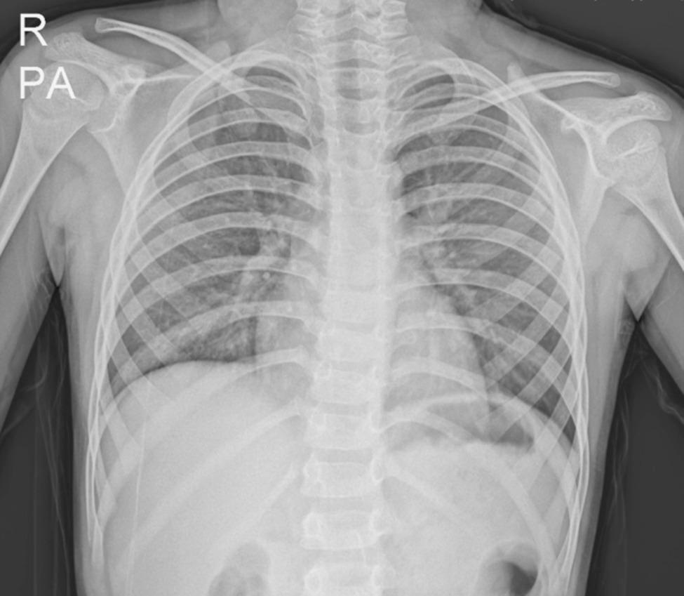
In paediatric patients the most likely source of CNS TB is from the pulmonary focus with hematogenous spread to the CNS. The meninges, the brain’s subpial or subependymal surfaces may be the site of the original Rich focus, which could lie dormant for many years. The precise stimulus required for the rupture and proliferation of these lesions is still not fully understood, however may be immunological in nature, depending upon the virulence of the bacteria and the immune resistance of the host [3, 4].
CNS TB can present as tubercular meningitis (TBM), focal or diffuse pachymeningitis, intracranial tuberculoma or brain abscess. Spinal TB can present as spinal meningitis and spinal arachnoiditis [3,4,5,6]. Spinal HSM is a rare complication of CNS TB. It is presumed to result from arachnoiditis-induced obstruction of CSF flow, leading to syrinx formation [6, 7]. Syrinxes are fluid-filled pockets that develop in the spinal cord, and this condition is called syringomyelia. Hydromyelia is described as the dilation of the central canal of the spinal cord. These two conditions are also known as HSM as they cannot be distinguished separately [8].
Neurological symptoms might range from agitation, lethargy to coma, depending on the stage of presentation. Patients with tuberculoma or tuberculous brain abscess may present with headache, convulsions, papilledema, or other symptoms of elevated intracranial pressure(3,4). In a spinal tuberculosis complicated by HSM, neurological symptoms can range in severity from minor discomfort to neurological impairments including sensory neural deficit and motor involvement [7].
Based on distinctive imaging and spectroscopic results, MRI can confidently diagnose tuberculomas. On T2W imaging, intracranial tuberculomas typically exhibit central hyperintensity or hypointensity with a hypointense rim, while on T1W images, they typically exhibit isointensity or hypointensity. Depending on central caseation, these granulomas exhibit either peripheral rim enhancement or homogeneous enhancement on fat suppressed T1W imaging following paramagnetic contrast injection. In vivo MRS can reveal decreased or absent NAA, choline, and creatine, as well as prominent lipid and lactate peaks [5, 9]. In TBM, imaging frequently reveals hydrocephalus, abnormal enhancement in the basal cisterns and acute ischaemic insult as a result of vasculitis [5, 9].
MRI characteristics of spinal meningitis and spinal arachnoiditis include matting of the nerve roots in the lumbar area, CSF loculation, and obliteration of the spinal subarachnoid space, together with a thinning of the spinal cord in the cervicothoracic spine. Arachnoiditis complications can include spinal cord involvement in the form of HSM and infarction [5, 7].
Based on the MRI findings and MRS results in the particular clinical scenario, which were backed by CSF results, we were able to confidently diagnosis tuberculomas, TBM and HSM in our case.
Although coexisting TBM and intramedullary or intracranial tuberculomas are reported in literature however a combination of tuberculomas, TBM, and Hydrosyringomyelia is a rare and very few cases are reported [7].
For children with severe ventriculomegaly, CSF diversion treatments must be carried out at the right time, particularly when patient has infracts as a complication, as in our patient. As far as tuberculomas in children are concerned, surgical intervention can be sought in patient with significant mass effect and in case with diagnostic dilemma to get histopathological insight. The need for surgical intervention has drastically reduced because of availability of effective antitubercular therapy (ATT); although multidrug resistance is a known challenge. Most lesions completely disappear with standard antitubercular treatment with measures to reduce cerebral oedema and mass effect using dexamethasone [10]. For HSM conservative treatment and high dose steroids suffice, use of intravenous immunoglobulin (IVIG) as part of the drug therapy with satisfactory outcome is documented [2].

