
PNP is a rare, autoimmune-mediated mucocutaneous disease that is almost always associated with an underlying neoplasm. The most common accompanying tumor is lymphoid tissue proliferative tumor, with Non-Hodgkin’s Lymphoma being the most common [6, 7], characterized by painful mucosal erosion and polymorphic skin lesions in the background of occult or confirmed tumors [8].
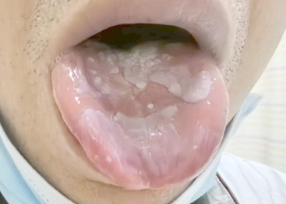
Mucosal involvement is considered to be a characteristic manifestation of PNP, usually manifested as severe oral mucositis, which may or may not be accompanied by various forms of skin lesions [2]. Histopathology typically shows acantholysis, lichenoid interface dermatitis, and keratinocyte necrosis with IgG and complement deposition in the interstitial spaces or basement membrane zones of the epidermis [9,10,11]. Lichen planus in paraneoplastic pemphigus/paraneoplastic autoimmune multiorgan syndrome (PNP/PAMS) is often associated with Castleman’s disease, a rare nonclonal lymphoid tumor that most commonly develops in the peritoneal cavity [12]. Studies have shown that such lesions may exist many years before the clinical manifestations of paraneoplastic autoimmune multi-organ syndrome appear, and have a long hidden course [13]. The patient in this case initially presented with oral lichen planus as the main symptom, and later came to our department due to coughing and wheezing after physical exertion. It took more than 4 months to be diagnosed with pulmonary small lymphocytic lymphoma. In this case, the patient’s extrapulmonary symptoms were limited to oral lichen planus, without typical skin lesions in other parts of the body. Considering that the oral lesions themselves are difficult to heal and affect eating, performing a tongue biopsy might have seriously impacted the patient’s quality of life due to delayed healing of ulcers and wounds. Therefore, following a discussion with the patient and his family, it was decided not to perform a tongue biopsy. Clinicians are advised to be alert to the possibility of paraneoplastic pemphigus and potential tumors in patients with oral mucosal diseases who do not respond well to conventional treatment.
BO is a small airway disease that primarily affects the terminal bronchioles, characterized by severe and irreversible obstructive ventilatory dysfunction. It is mainly caused by factors such as organ transplant rejection, connective tissue diseases, viral infections, inhalation of toxic substances, and autoimmune disorders [14,15,16]. BO is a serious complication characterized by dry cough, dyspnea, and hypoxemia, affecting approximately 30–90% of PNP patients [13, 17]. On CT imaging, it may present as “mosaic” perfusion, bronchial wall thickening, centrilobular nodules, and bronchiectasis [18]. Research has found that respiratory failure caused by BO is the main cause of death in PNP patients with Castleman’s disease [13]. When PNP is complicated with BO, the patient’s risk of death significantly increases, often indicating poor prognosis [3].
This patient initially presented with oral lichen planus. He denied any history of tobacco use, chronic respiratory illness, or work-related inhalation of dust. He presented with dry cough, followed by wheezing after physical activity. Blood gas analysis showed hypoxemia. Immunological screening tests were unremarkable. Pulmonary function tests showed extremely severe mixed ventilation dysfunction. Chest imaging revealed thickening of peripheral bronchiolar walls, central nodules in the lobule, and partial bronchiectasis in both lower lobes of the lungs. Areas of decreased lung attenuation were also observed, indicating air trapping—a hallmark radiological feature of small airway disease. Based on these clinical, functional, and radiological findings, a definitive diagnosis of bronchiolitis obliterans (BO) was established. Combined with the characteristics of the patient’s pemphigus, positive anti-desmoglein 3 antibodies, and the final mediastinal puncture biopsy confirmed the diagnosis of pulmonary small lymphocytic lymphoma.
Lymphoma, PNP, and BO can constitute a clinical syndrome. Its exact pathogenesis is not yet clear, but it is speculated that certain autoantibodies produced by PNP may play a role in causing acute inflammation of the respiratory epithelium [4, 5]. Several patients have PNP onset, which may be difficult to detect early due to the insidious onset of lymphoma and BO. Some lymphoma patients may develop PNP months or years later [19]. In these patients, lymphomas often progress slowly and are of low-grade malignancy, but their prognosis is poor, with respiratory failure caused by BO being the main cause of death. Therefore, patients with PNP or unexplained respiratory symptoms should be highly alert to the possibility of potential lymphoma and undergo a comprehensive evaluation to achieve early diagnosis and intervention.
A multicenter study in China showed that for patients with chronic lymphocytic leukemia/small lymphocytic lymphoma who were > 65 years old or had high-risk factors, doctors preferred a regimen containing a BTKi, followed by a regimen containing rituximab; for patients who were ≤ 65 years old and without high-risk factors, the proportion of patients who chose the two regimens was similar [20]. Patients with BO have a poor prognosis. Current therapeutic strategies primarily aim to alleviate symptoms, delay disease progression, and prevent complications. Conventional pharmacological treatments include corticosteroids, immunosuppressive agents, and antibiotics. Additionally, supportive therapies such as long-term oxygen therapy and pulmonary rehabilitation play an important role. In specific circumstances, lung transplantation can be used as the final treatment option for severe patients who are ineffective with drug treatment [21]. This patient received standard inhaled corticosteroid treatment, combined with two courses of Orelabrutinib combined with Obinutuzumab to treat the primary disease. The patient’s lymphoma and paraneoplastic syndrome improved, but subsequent chest CT examination still showed imaging manifestations consistent with BO.
The limitation of this case is that no pathological evidence was obtained for BO, mainly because the patient had a mediastinal tumor, bronchoscopic biopsy was limited, and the patient had poor lung function and no bronchoscopy was performed.