
A 518-million-year-old fossil has revealed that some of the earliest vertebrates possessed four image-forming eyes instead of two.
That configuration recasts a small brain structure humans still carry as the remnant of a once fully visual organ.

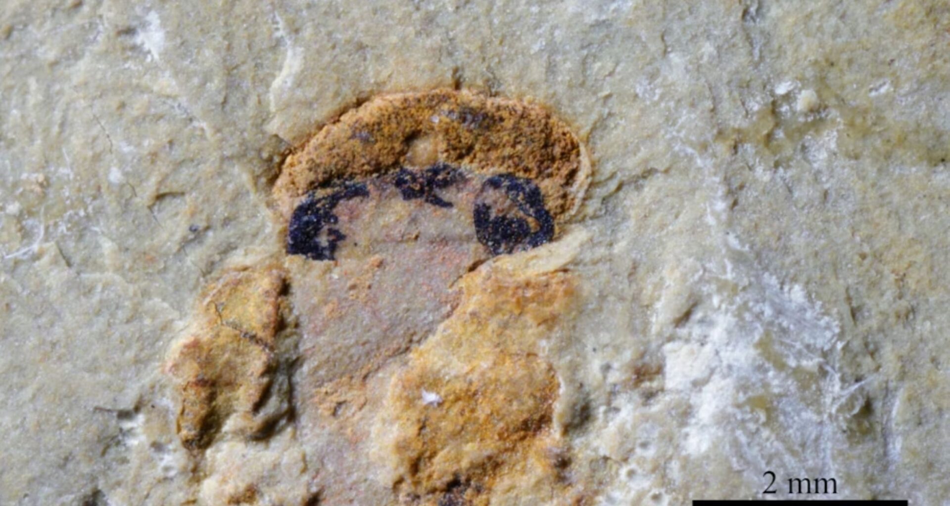
Embedded in rock from southern China, the head of Myllokunmingia, an early jawless fish that lived more than 500 million years ago, preserves two large lateral eyes and two smaller organs aligned along the midline.
Evidence in stone
Examining those central structures, Prof. Peiyun Cong at Yunnan University documented pigment patterns and anatomy consistent with functioning eyes rather than random discoloration.
Both midline organs occupy the precise position later vertebrates reserve for the pineal system, tying this ancient four-eyed layout to a feature that survives deep within our own brains.
That placement leaves open a larger question about how and why a fully visual organ was reduced over time, a shift that requires closer examination of the tissue itself.
Rare fossils preserve eyes
Fine mud at the Chengjiang site in Yunnan, southern China, sometimes locked soft tissues in place.
Rapid burial cut off oxygen, slowed bacteria, and gave minerals time to copy delicate structures before they collapsed.
High-powered microscopes sharpened the view of those patches, letting researchers track each eye spot through the rock.
That kind of preservation turns a smudge into a body part, making chemical clues possible in the first place.
Chemistry behind dark spots
Chemical tests checked whether the spots carried the same pigments that make modern eyes dark and light-sensitive.
Using spectroscopy, a method that reads how materials absorb light, the team compared the spot signals with living eye tissue.
Results pointed to melanosomes, tiny pigment packets inside cells, matching the shape and density found in the animal’s larger eyes.
With those pigmented cells in place, the smaller central organs likely absorbed light, supporting the idea that they formed images.
A pair in center
Between the side eyes, the team identified two midline organs that matched where many backbone-bearing animals carry a pineal system.
In living animals, the pineal complex – paired organs near the brain’s roof – grows from the same early tissue as eyes.
Instead of a simple light meter, those organs carried pigment and lens-like parts, tying them to vision.
That link suggests the pineal system did not begin as a sleep aid, but as extra eyes.
Evidence for image formation
Lens-shaped structures showed that each organ did more than sense brightness, because a lens can focus an image.
Scientists call these camera-type eyes, eyes that use a lens to make images, and the fossils fit that plan.
Having a second image-forming pair on top could have widened what the animal noticed without turning its head.
No fossil can show sharpness or color, so the exact quality of those images remains out of reach.
Survival in early seas
During the Cambrian Period, an early time when animal diversity grew fast, predators pushed small swimmers to spot danger sooner.
Myllokunmingids belonged to the first known vertebrates, animals built around a backbone and spinal cord.
Extra eyes could have improved predator detection from above, where shadows often arrive first in shallow water.
Still, the fossil does not sit on the human family line, so its vision shows options, not guarantees.
From eyes to glands
Later vertebrates kept the midline organs but reduced their job, turning them into light sensors rather than full eyes.
Loss of a working lens meant less visual detail, while deeper placement protected the tissue and cut the cost of upkeep.
“Only later in evolution did they shrink, lose visual power and take on their modern role in regulating sleep,” Cong said.
Today that reduced system still responds to light, but many species use it mainly to time body chemistry.
The pineal gland today
Deep in the human brain, the pineal gland, a small organ that reacts to light, now sits far from the eyes.
At night it releases melatonin, a sleep-linked hormone that signals darkness to the body, and that signal helps start sleep.
That timing supports your circadian rhythm, the roughly 24-hour cycle that sets sleep and alertness across each day.
Disruptions in light exposure can confuse that clock, yet the pineal gland does not form images the way eyes do.
Limits and open ends
Only two myllokunmingid species showed the extra organs, so the fossil record cannot yet map how common that layout was.
Hard parts like lenses leave clearer traces than nerves, so the pathway from those organs into the brain stays hidden.
To test whether this four-eye plan was ancestral, researchers will need other early vertebrate fossils with equally preserved heads.
Until then, the finding stands as a rare case where soft tissue rewrites what a backbone animal looked like.
What this changes
Seen in one rare fossil, extra image-forming organs connect ancient eyesight to a modern brain gland, tightening a long suspected link.
Future finds from the same beds could show when that visual hardware faded, and which living lineages kept any version of it.
The study is published in Nature.
—–
Like what you read? Subscribe to our newsletter for engaging articles, exclusive content, and the latest updates.
Check us out on EarthSnap, a free app brought to you by Eric Ralls and Earth.com.
—–