
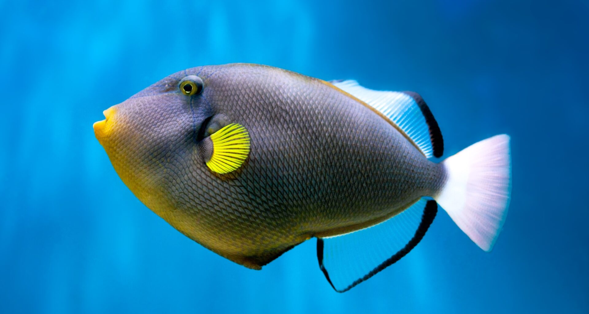
The inside of a fish’s skull has long seemed like a simple story. Scientists assumed that the hollow space left behind after soft tissue decay could act as a reliable stand-in for the brain.
This idea shaped decades of research on how vertebrate brains evolved. A new study now challenges that assumption and reveals a far more complex picture.
Looking inside fish skulls

For years, researchers relied on endocasts, the natural fills of skull cavities, to study ancient brains.
These casts often preserve the shape of the space where the brain once sat. Scientists treated them as close replicas of real brain structures.
This method worked well in many vertebrates. In mammals and birds, the brain fits tightly within the skull. The cavity reflects the brain’s shape with reasonable accuracy. Fish, however, tell a different story.
A major knowledge gap
Ray-finned fishes make up about half of all vertebrate species. Despite this, scientists knew very little about how their brain cavities compare to their actual brains. Only a few species had been studied in detail.
This gap raised an important question. If the relationship between brain and cavity differs in living fish, then fossil interpretations may also be flawed.
“How can we pretend that we understand how evolution, and especially brain evolution, works if we know almost nothing about half of the vertebrates?” asked Rodrigo Figueroa, first author of the study.
Scanning fish in detail
To answer this, researchers examined 86 species of ray-finned fishes. These species covered a wide range of habitats and evolutionary branches. The team used a technique called dice-CT scanning.
They soaked the fish in iodine, which makes soft tissues visible in scans. This allowed them to see both the brain and the surrounding cavity in three dimensions without cutting the specimen.
The result was a detailed look at how brains actually sit inside skulls.
No common structure pattern
The findings surprised the researchers. In many vertebrates, endocasts follow a clear and consistent pattern. Scientists can identify regions like the optic lobes or hindbrain across species.
Ray-finned fishes break this pattern. Some species show clear internal divisions in their cavities. Others have smooth, simple shapes with no obvious features. Even closely related species can look very different.
“If you CT scanned a mammal skull and made a replica of the brain by filling in the empty space inside, it would look very similar to the actual brain; there is a very tight fit between the brain and skull,” said Stephanie Pierce from Harvard University.
“But, with these fishes, there’s small brains, big brains, smooth brains, convoluted brains; it’s just astonishing the amount of diversity these animals are showing.”
Brain does not fill space
The study also measured how much of the skull cavity the brain actually occupies. This is called the brain-to-endocranial cavity ratio.
In birds and reptiles, the brain usually fills most of the cavity. In ray-finned fishes, the average is much lower. In some species, the brain occupies less than 5 percent of the space.
In others, it fills more than half. This wide range shows that the brain often sits within large empty areas.
Deep sea extremes
Habitat plays a strong role in this pattern. Deep sea fishes tend to have very low brain-to-cavity ratios. Their brains are small compared to their skull size.
This may relate to energy use. Brain tissue requires a lot of energy, and food is limited in deep ocean environments. A smaller brain may reduce energy demands.
“Many things can explain why it would be beneficial to have a tiny brain and a big head,” Figueroa said. “The meningeal tissue surrounding the tiny brain acts as a protective bumper that provides a space for the brain, keeping it safe from impact or variations in overwhelming pressure.”
Skull shape influences cavity
The shape of the skull also affects the cavity. Flattened skulls tend to produce flattened cavities. Elongated skulls create elongated spaces.
However, this relationship is not always straightforward. Some predators have skull shapes that prioritize feeding over brain placement. This leads to unusual cavity shapes that do not match expectations.
Other structures, like jaw muscles and inner ear organs, also compete for space inside the head.
Growth changes brain fit
The study looked at different life stages in a species called Amia. Young fish have brains that fill more of the cavity. As they grow, the skull expands faster than the brain.
This creates more empty space in adults. It shows that age can affect the brain-to-cavity relationship.
This finding adds another layer of complexity when studying fossils, where the age of the specimen is often unknown.
Rethinking fossil brains
These results have major implications for paleontology. Scientists often describe fossil endocasts as if they directly represent brain anatomy.
The study shows that this approach can be misleading, especially in ray-finned fishes. The cavity does not always match the brain’s shape or size.
“For decades researchers assumed a fossil’s endocast shape directly reflected its brain morphology, and while that’s a pretty good assumption for most vertebrates, it isn’t for fish,” Pierce said.
“By comparing endocasts to brains, our findings show that the two can evolve on completely different trajectories.”
Instead of acting as brain replicas, endocasts may reflect other factors. They can reveal information about habitat, skull mechanics, and sensory systems.
This shifts their role in research. Scientists may use them to study ecological adaptations rather than brain structure alone.
Future research directions
This work opens new questions rather than closing them. Researchers may find clearer patterns within smaller groups of fishes. More data could reveal links between brain shape, environment, and behavior.
“Modern neuroscience often focuses on mapping the ‘connectomes’ of a few specific species,” Figueroa noted.
“Yet this study shows that such a focus provides only a small, narrow view of what has actually evolved over millions of years and it raises more questions about whether the flexible, diverse brain shapes of these fishes caused their global success, or whether their success in varied environments forced their brains to adapt in such unique ways.”
A shifting perspective
This study changes how scientists view the fish skull. What once seemed like a simple cast of the brain is now a complex structure shaped by many factors.
“This study represents our first steps,” Pierce added. “It’s giving us a quick snapshot of what the major trends are, and where we go next.”
The message is clear. The space inside a fish’s skull holds more than just clues about its brain. It carries traces of evolution, environment, and survival strategies across millions of years.
The study is published in the journal Proceedings of the Royal Society B Biological Sciences.
—–
Like what you read? Subscribe to our newsletter for engaging articles, exclusive content, and the latest updates.
Check us out on EarthSnap, a free app brought to you by Eric Ralls and Earth.com.
—–