
In total, 27,171 new transcripts, associated with 24,715 genes, were identified after accounting for multiple transcripts derived from the same gene. The analysis yielded expression data for 24,715 genes across 14 tissues in our transcriptome. Among them, there are a total of 70 genes were related to the innate immune pathway.
Gene Ontoloty analyse
According to the results of differential gene detection, GO analysis divided DEGs into three functional categories: molecular function, cellular components, and biological processes (Fig. 2).
Fig. 2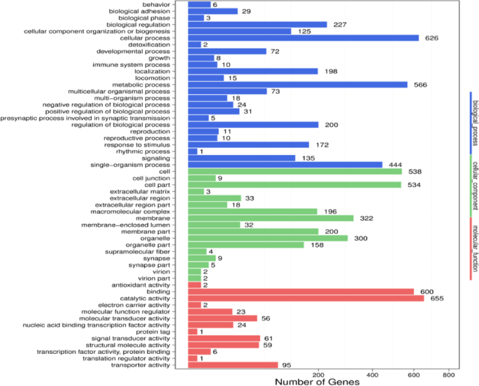
GO functional classification of DEGs.
Pathway function analysis of DEGs
Based on the testing of DEGs, we performed pathway classification of the DEGs (Fig. 3b) and the enrichment analysis of KEGG pathways (Fig. 4b)20,21,22,23.
Fig. 3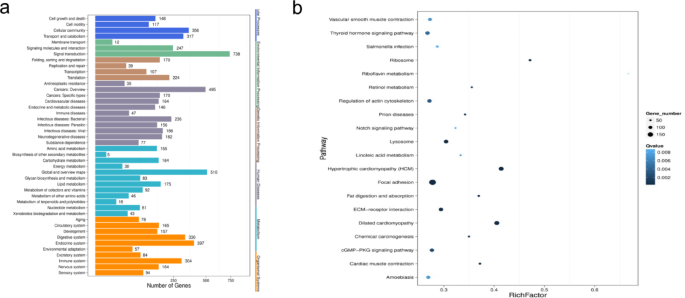
Pathway classification of the DEGs (a) and enrichment analysis of KEGG pathways (b).
Fig. 4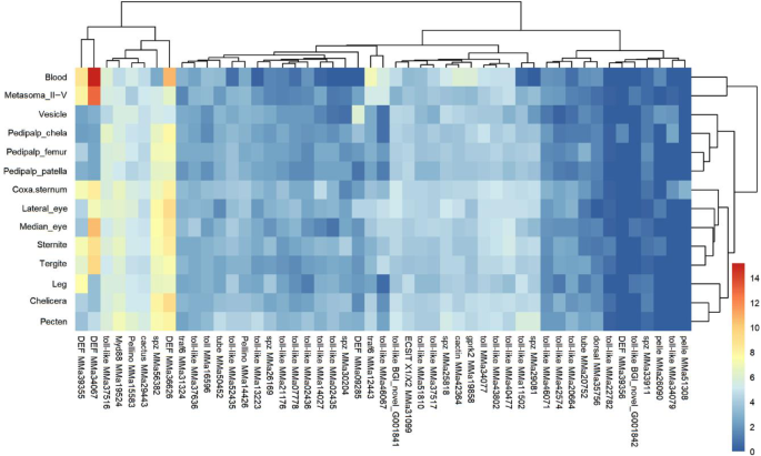
Heat map showing the expression levels of various Toll pathway-related genes across 14 Mesobuthus martensii tissues. The horizontal axis of the heat map represents the screened differentially expressed genes, while the vertical axis corresponds to the 14 tissue samples. The color gradient also reflects the level of gene expression, with red and blue colors indicating high and low expression, respectively.
For Fig. 3a, the X-axis represents the number of DEGs, and the Y-axis represents the KEGG pathways. For Fig. 3b, the X-axis represents enrichment factor values, and the Y-axis represents name of pathways. Color represents qvalue (the white color means higher value, and the blue color means lower value), and while the value smaller, the enrichment result is more significant. The size of the dots represents the number of DEGs (larger dots represent larger numbers, smaller dots represent fewer numbers).
Microbial recognition genes
The immune genes of four pathways derived from the KEGG pathway (https://www.genome.jp/kegg/pathway). First, we found the immune genes of four pathways and receptors. Then, we searched for the annotation of our RNAseq to sift the potential genes. Finally, we identified those genes by using BLAST. Also, we made a systematic BLAST search analysis based on some other invertebrates to discuss the diversity of immune genes and pathways among different invertebrates (Table 1). Among the innate immune genes, the expression in blood was the most unique.
Table1 Comparison of the number of immune genes in four arachnids, four insects, and two crustaceans.Lectins (lectins)
In M. martensii, the following five galectin-expressing genes were identified: BGI_novel_T010761 which exhibited non-specific expression across tissues; MMa18050 which had a low expression in blood (Fragments Per Kilobase of exon per Million fragments mapped (FPKM) = 14.72) but highly expressed in venom vesicles (FPKM = 548.94); MMa18049 was exclusively expressed at low levels in blood and MMa50712 expressed at low levels in blood; and MMa51092 which displayed very low expression to no expression in all tissues (FPKM not exceeding 6).
Down syndrome cell adhesion molecule (Dscam)
M. martensii’s transcriptome revealed at least 95 potential transcripts of Dscam arising from variable splicing, with most of them exhibiting low expression levels and no clear tissue-specific patterns.
The Nimrod superfamily
In M. martensii’s transcriptome, only one homologous gene to draper was identified, with its expression levels showed no significant variations across different tissues.
Innate immune signal transduction pathwaysToll pathway
The transcriptome of M. martensii revealed one Dorsal, one Cactin, one Cactus, and two TUBE genes. Subsequent heat map and hierarchical clustering analyses of gene expression in the Toll pathway indicated that most of these genes were expressed at low levels under innate conditions (Fig. 4). Specifically, the expression levels of spatzle (spz) genes (MMa25818 and MMa26169) relatively average among tissues, while those of genes (MMa29081 and MMa30204) were undetectable in blood (Supplementary Material 1). Conversely, (MMa56382) showed a low expression level of 5.1 in blood tissues, with its expression level in vesicles and metasoma II-V were also low, thus differing from those observed in other tissues (Supplementary Material 1). Overall, spz genes exhibited low expression levels in blood, with results for (MMa29081 and MMa30204) aligning with those of previous studies on Macrobrachium rosenbergii24. Multiple Toll and TLR (toll-like receptor) genes were also identified in M. martensii’s transcriptome, most of which were expressed at low levels and lacked significant tissue-specific differences. Upon activation, Toll binds to the adaptor protein MyD88 and recruits the interacting proteins Tube and the kinase Pelle, thereby forming the MyD88-Tube-Pelle heterotrimeric complex25. Expression analysis further revealed no significant tissue-specific patterns for MyD88 and Tube in the transcriptome of M. martensii. However, on average, Tube’s expression level was lower than that of MyD88, while the expression of Pelle was even lower than that of both MyD88 and Tube. Notably, Pelle (MMa26090) exhibited low expression in blood, lateral eyes, ventral plate and vesicles (FPKM = 2.11, 1.18, 1.16 and 3.18 respectively), while Pelle (MMa51308) was exclusively expressed in the coxa-sternum. Additionally, Pellino, a positive regulator of the Toll signaling pathway26, did not exhibit significant tissue-specific expression patterns.
Cactus acts as a repressor by inhibiting the NF-kB transcription factor Dorsal when the Toll pathway is inactive, but upon the formation of the MyD88-Tube-Pelle complex, it undergoes phosphorylation, mediated by Pelle, and subsequent degradation27,28. Once activated, Dorsal translocates to the nucleus where it binds to the kB-related sequence of the antimicrobial peptides (AMP) gene29. In the transcriptome of M. martensii, neither Cactus nor Dorsal genes exhibited tissue-specific expression. However, Dorsal’s expression level was significantly lower than that of Cactus, thus reflecting differences in the Toll pathway’s function in scorpions compared with insects (Fig. 5).
Fig. 5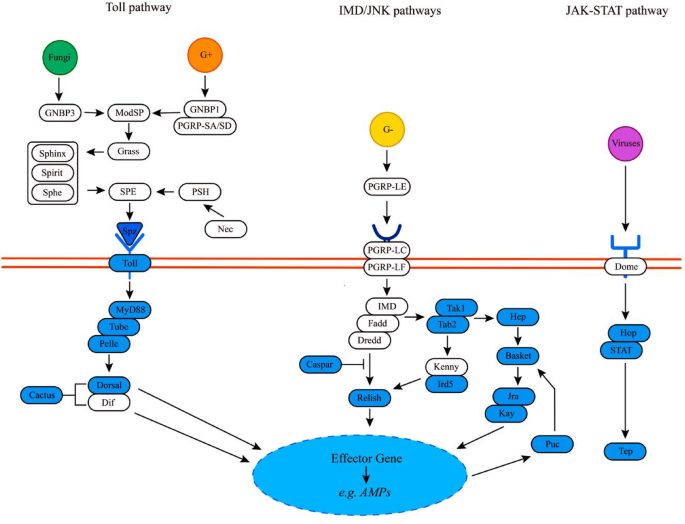
The four primary immune pathways in Mesobuthus martensii: Toll, IMD, JNK, and JAK/STAT. These pathways responded to various immune challenges and ultimately leading to the transcriptional activation of genes involved in phagocytosis, encapsulation and humoral responses. Genes identified as part of the immune signaling pathways of M. martensii are highlighted in blue, while unverified ones are depicted in white.
IMD pathway
In insects, the IMD signaling pathway plays a crucial role in defending against gram-negative bacteria, and it exhibits similarities to the TLR pathway in mammals30. In this study, transcriptome analysis of M. martensii revealed the presence of two Tak1 genes, one Tab2 gene, two Iap2 genes, one Ubc13 gene, and two Uev1a genes. However, the Effete (Ubc5) gene was not identified. Both Tak1 and Tab2 genes showed non-tissue-specific expression patterns, and among the Tak1 genes, one Tak1 (MMa52113) exhibited lower expression levels than another Tak1 (MMa37335). Furthermore, the Ubc13 (MMa14412) gene did not display significant tissue-specific expression compared with the Uev1a (MMa12278) and Uev1a (MMa44597) genes.
The Kenny-Ird5 kinase complex, along with Dredd, facilitates the cleavage of Relish, but Casper can inhibit this Dredd-mediated cleavage process31. Interestingly, M. martensii lacked the Kenny gene but possessed one Ird5 gene, with the latter being ubiquitously expressed across tissues but showed a relatively lower expression level in the leg (FPKM=1.17). Additionally, the transcriptome of M. martensii contained two Relish genes which exhibited higher expression levels in the pectens as well as three Casper genes which did not show distinct tissue-specific expression patterns (Fig. 6).
Fig. 6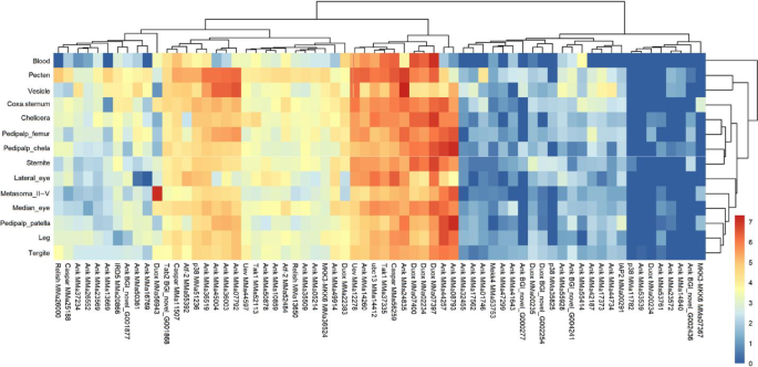
Heat map representation of the expression levels of various IMD pathway-related genes across 14 Mesobuthus martensii tissues.
The IMD pathway primarily responds to Diaminopimelic acid peptidoglycans in gram-negative bacteria32. Upon binding to the transmembrane PGRP, the DAP peptidoglycan activates the pathway which subsequently interacts with cytoplasmic PGRP and IMD to initiate the signaling cascade. Then, IMD associates with Fadd and recruits Dredd33, leading to its modification and the subsequent activation of TAK1 and TAB234. The Kenny-Ird5 kinase complex is phosphorylated by Tak1 and Tab2, and alongside Fadd and Dredd, this complex co-activates the NF-kB transcription factor Relish35. After cleaving the Ankyrin repeat sequence, the remaining Relish fragment translocates to the nucleus and works with other effectors to induce AMP expression36. In Drosophila melanogaster, the E3-ligase inhibitor of apoptosis 2 (Iap2) is activated by Dredd activity, facilitating the recruitment and activation of the Tab2/Tak1 complex37. This process is further supported by E2 ubiquitin-conjugating enzymes such as Uev1a, bendin (Ubc13), and Effete (Ubc5)38. In the transcriptome of M. martensii, the FPKM values of IMD, Fadd, and Dredd genes were 0. By comparison, in Drosophila melanogaster, the expression of these three key genes is essential for initiating the transcription of downstream genes.
JNK pathway
The c-Jun N-terminal kinases (JNKs), part of the mitogen-activated protein kinase (MAPK) family, are involved in various biological processes, including damage repair, cellular metabolism, apoptosis, and tissue homeostasis39. The composition of the JNK pathway in M. martensii resembled that of Drosophila melanogaster40 and Anopheles gambiae41, with the transcriptome analysis revealing the presence of one JNKK (Hep) gene, four JNK (Basket) genes, two Jra (c-Jun) genes, one Kay gene, and three Puc genes. The MAPK kinase Hep genes exhibited low expression levels in the blood, with similar results were observed in the JNK genes such as Bsk (BGI_novel_G002151), Bsk (MMa30726), and Bsk (MMa45799). However, both Jra and Kay genes showed higher expression levels than Bsk and Hep, although their expression in blood was still low. On the other hand, while the Puc gene also exhibited a relatively low expression level in the blood, it was less pronounced than the other JNK pathway genes. Overall, the JNK pathway in M. martensii demonstrated a generally low expression level in blood tissues. In contrast, the Kay gene displayed a higher level of expression in specific tissues compared with other genes within the JNK pathway (Fig. 7).
Fig. 7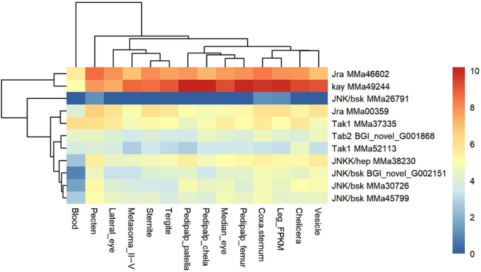
Heat map showing the expression levels of various JNK pathway-related genes across 14 Mesobuthus martensii tissues.
The JNK pathway is critical for regulating humoral immunity and immune-mediated epithelial shedding in insects42. Additionally, it plays a significant role in antiparasitic defense mechanisms in Drosophila melanogaster43. As an integral component of the IMD-associated pathway, the JNK pathway can be activated through the induction of MAPK kinase kinase (Tak1) and Tab2 binding in Drosophila melanogaster44. However, current understanding of the JNK pathway in arthropods, including scorpions, remains incomplete and requires further experimental validation45.
JAK-STAT pathway
The JAK-STAT pathway is mainly recognized for its role in virus-induced signaling cascades, but it is also involved in regulating cell growth, differentiation, apoptosis, and inflammatory responses46. In Drosophila melanogaster, three types of cytokine unpaired molecules (Upd1-3) bind to the transmembrane receptor Dome to initiate the JAK/STAT pathway47. Subsequently, Dome transmits signals to Hop and STAT92E to activate the transcription of related genes48.
In M. martensii’s transcriptome, Upd, Dome, and some regulatory factors are absent. However, one Hop, one STAT, four inhibitors of cytokine signaling (SOCS), and one protein inhibitor of activated STAT (PIAS) were identified. Among these, Hop (MMa32976) did not exhibit significant tissue-specific expression, while STAT5B (MMa12827), for which the expression level was low, was not expressed in the blood. Similarly, PIAS2 (MMa45841) was not expressed in the blood, mid-eye and ventral plate, but exhibited low expression levels in other tissues. Furthermore, SOCS2 (MMa36355) was expressed at a low level in the vesicle (FPKM = 46.74), while the expression of SOCS7 (MMa52300) was significantly lower than the other three SOCS genes and lacked significant tissue specificity. Finally, SOCS (MMa39073) and SOCS (MMa54882) did not display any distinct tissue-specific expression patterns. Overall, the expression of JAK-STAT pathway genes was relatively low compared with their negative regulators, hence suggesting that the pathway was suppressed under innate conditions in M. martensii.
In arthropods, infection triggers an innate immune response which involves four main signaling pathways (Toll, IMD, JNK, and JAK/STAT) that collectively activate antimicrobial defense mechanisms. Scorpions, which are constantly exposed to microbes in their habitats, exhibit both inducible and non-inducible immune response mechanisms, with such responses being evident in their venom glands and hemolymph when exposed to Escherichia coli, Micrococcus luteus or infectious injuries.
EffectorsAntimicrobial peptides (AMPs)
Defensins: To date, six known defensins49 and two putative ones50 have been identified in scorpions, but in this study, a novel defensin gene MMa34067 (BmKDfsin9) was hypothesized. Additionally, the expression of BmKDfsin1, BmKDfsin2, BmKDfsin4, and BmKDfsin6 genes was not detected in the transcriptome of M. martensii.
Furthermore, BmKDfin3 was expressed at low levels only in the pedipalp chela, while BmKDfsin5 was expressed in all tissues, with relatively low levels in the vesicle but significantly high expression in the blood (FPKM = 1677.64). Conversely, the average expression of BmKDfsin7 was much lower than that of BmKDfsin5, and it was not expressed in blood. However, BmKDfsin7 also showed higher expression in the venom sacs than in other tissues. In the case of BmKDfsin8, it exhibited a similar expression pattern to BmKDfsin5, although it was lower expressed in some tissues. Finally, BmKDfsin9 displayed a high expression level in blood (FPKM = 37,038.68), significantly higher than other tissues, with metasoma II-Ⅴ having the second-highest expression (FPKM = 7768.4).
Despite the absence of genes upstream of the IMD pathway and the generally low expression of downstream genes in M. martensii, certain defensin genes (BmKDfsin5, BmKDfsin8, and BmKDfsin9) showed high expression levels in blood, hence highlighting a potentially constitutive role in immune defense.
Antimicrobial peptides in venom: Using the NCBI database and M. martensii transcriptome data, two venom AMP genes, namely BmKb1 and BmKa2, were identified, both of encoding proteins belonged to the same group of disulfide-free peptides and shared similar expression patterns. These genes were also highly expressed in the vesicle and posterior metasoma II-Ⅴ, with minor expression in the steppe, cheliped and antennal limb. However, BmKbpp, BmKn2, and Marcin18, along with their homologous genes,49 were not detected in the transcriptome data.
For further analysis, we used local BLAST comparisons of venom AMP sequences across various scorpion species to reveal numerous homologous genes, many of which were annotated as venom peptides. This high homology not only underscores the close relationship between AMPs and venom peptides but also suggests that AMPs may protect venom glands from infection. Meanwhile, it also may enhance the efficacy of neurotoxins51.
Lysozyme (Lysozyme)
In the transcriptome of M. martensii, 15 lysozyme family genes were identified. Among these genes, Lysozyme (BGI_novel_G002825) exhibited the highest expression in the coxa-sternum, followed by high expression in the chelicera but low expression in other tissues. Conversely, Lysozyme (BGI_novel_G003726) was highly expressed in the vesicle, with secondary expression in the coxa-sternum, while other tissues expressed at lower levels. Furthermore, Lysozyme (MMa18052) was predominantly expressed in the blood, with the lowest expression in the vesicle and low levels also observed in other tissues. Similarly, Lysozyme (MMa33840) had the highest expression in the blood, with significant high expression also occurring in the lateral eye, middle eye, metasoma II-V, sternite, and tergite. In the case of Lysozyme (MMa31766), no expression was detected in the blood, while its expression levels were low in other tissues. Finally, Lysozyme (MMa42986) was highly expressed in the lateral eye and vesicle, with no expression in the pectens and pedipalp (chela, femur, and patella), while Lysozyme (MMa44672) exhibited the highest expression in the lateral eye, with average lower levels in other tissues. The remaining genes had low expression or no significant differences in expression.
Thioester-containing proteins (TEPs)
TEPs were historically believed to be synthesized via the JAK/STAT pathway, thereby indirectly contribute to immunity by tagging pathogens for phagocytosis52. The CD109 antigen, a member of the TEP superfamily, exhibits multiple anchoring functions and plays a key role in cellular immunity, as demonstrated in the cotton bollworm where it is induced by the ecdysone pathway53,54,55.
Previous research identified two TEP genes in M. martensii19, but the expanded analysis undertaken in this study revealed 16 homologous CD109 antigen genes in M. martensii’s transcriptome. This discrepancy may resulted from some genes originating from distinct transcripts. Among those identified genes, TEPs (MMA25797) exhibited high expression levels in the blood, whereas the remaining CD109 antigen homologues displayed low or undetectable expression in blood. Additionally, most of the CD109 homologues exhibited high expression levels in the pectens.