
Chemicals and equipment
All media ingredients, reagents, and chemicals were purchased as follows: Methanol (Th. Geyer, Renningen); water for analytical and preparative HPLC (VWR International GmbH, Dresden); formic acid (VWR International GmbH, Dresden); acetonitrile (Th. Geyer, Renningen); Diaion HP20 resin (>250 µm beads; Thermo Scientific); DMSO-d6 (Sigma Aldrich, USA); rifampicin (TCI, Germany); media ingredients (Supplementary Table 1; Carl Roth, Germany). The HRMS data were collected on the Orbitrap Exploris 120 Mass Spectrometer (Thermo Fisher Scientific) equipped with an electrospray ion (ESI) source and a Vanquish Core UHPLC system (Thermo Fisher Scientific). High-performance liquid chromatography (HPLC) was carried out by using a Vanquish Core UHPLC system (Thermo Fisher Scientific). The chromatography conditions are indicated in the respective sections below. The NMR spectra of the peptidic compounds were collected using NMR spectrometers outfitted with Bruker CryoPlatforms (500 and 700 MHz).
Genome miningBiosynthetic repertoire
To identify strains potentially capable of producing demiguisins with higher yields, partial sequences (dmgP7, dmgP1, and dmgP1 to dmgP7) of the BGC responsible for producing demiguisin (dmg) [location: 317,893–327,300; total length: 9408 nt, region 3.2] from the genome of Amycolatopsis sp. PS_44_ISF1 was subjected to nBLAST analysis on NCBI5. Genomes of strains showing ≥85% sequence identity with the partial NRPS BGC were retrieved and analysed with antiSMASH v7.1 using bgcFlow wrapper 0.4.0 (conda environment) using default parameters10,29,30. In addition to antiSMASH, the genomes of both strains were additionally submitted to PRISM 4, and the substrate specificity of the adenylation domains in dmg and dem BGCs was predicted using PARAS and PRISM 422,23. BGC similarity networks were constructed with the antiSMASH output results for all strains using BiG-SCAPE v2.0 with the parameters –include_singletons –mix –mibig –version 4.011. The default cutoff value of 0.3 was applied. Networks were visualized in Cytoscape v3.8.1 (https://cytoscape.org/) and manually curated18. The dmg BGCs were further examined using Clinker of CompArative GEne Cluster Analysis Toolbox (CAGECAT) using the default parameters (min. alignment sequence identity of 0.3), and homologous gene clusters were visualized with Clustermap.js14.
Mining for kerA and kerC genes
To assess if both Amycolatopsis strains encode for keratinases, the KerA protein sequence (GenBank ID: AAB34259.1) and KerC protein sequence (GenBank ID: ABY65903.1) were used in BLASTp searches against the PS_44_ISF1 and NBC_01488 genomes20. The top hits from these searches were tabulated and compared for sequence identity, high query coverage, and E-values.
Taxonomic classification
To refine taxonomic placement of the strains containing the dmg BGC, genome FASTA files were subsequently uploaded to the Type Strain Genome Server (TYGS) for whole-genome-based phylogenomic analysis7. The resulting phylogenomic tree was exported in Newick format and visualized using iTOL (Interactive Tree of Life) v7.2.1 (https://itol.embl.de/)31. Finally, FastANI was performed using bgcFlow wrapper 0.4.0 to investigate species-level relatedness by calculating pairwise average nucleotide identity (ANI) between the genomes of strains incorporated into the TYGS analysis as the closest related reference strains to PS_44_ISF1 and NBC_014889,29.
Strain cultivationMedia screening
Three media were selected to optimize production of various secondary metabolites (Supplementary Table 1), including PDA, peptone dextrose broth (PDB), and a minimal broth (M9) supplemented with glucose and casamino acids. Seed cultures of Amycolatopsis sp. PS_44_ISF1 and Amycolatopsis sp. NBC_01488 were prepared in 25 mL of PDB in a 100 mL Erlenmeyer flask and cultivated for three days at 30 °C with shaking at 150 rpm. For PDB and M9 broth cultures, 100 µL of seed culture was used to inoculate 100 mL of PDB or M9 broth (1 mL of seed culture was centrifuged and resuspended in M9 broth prior to inoculation) in 300 mL Erlenmeyer flasks, in triplicate. The flasks were incubated at 30 °C for 14 days with shaking at 180 rpm.
For broth cultures, the culture was transferred to 50 mL falcon tubes, centrifuged at 10,000 rpm for 10 min, and the cell free supernatant was transferred into a Schott bottle containing 20 g/L of MeOH-activated Diaion HP20 resin and left overnight at room temperature. The beads were filtered, rinsed with 10% MeOH, and eluted with 50% and 100% MeOH until the eluent was colourless. The eluent was poured off into round bottom flasks and dried in vacuo. The dried extracts from PDB and M9 were then dissolved in 10% MeOH and partially purified using C18 solid phase extraction (SPE). The SPE column was first pre-activated (100% MeOH) and equilibrated (10% MeOH) column, where after the crude extracts were loaded onto the column. The column was washed with 10% MeOH, and the metabolites were eluted with 50 and 100% MeOH. Samples were pooled and dried in vacuo, and weighed for LC-HRMS/MS analysis. In all cases, media controls were included.
For agar cultures, 100 µl of seed culture was used to spread plate onto 90 mm PDA plates in triplicate, and were incubated at 30 °C for 14 days. Following incubation, the agar was cut into small pieces (1 × 1 cm), and the metabolites were extracted in a beaker with MeOH overnight. Afterwards, the solvent was filtered off (using filter paper) and evaporated in vacuo. The dried PDA extracts were then dissolved in 10% MeOH and partially purified using C18 solid phase extraction (SPE) as described above.
Eco-mimetic cultivations
Two media, containing either pheasant feathers or feather meal (Supplementary Table 1), were selected to investigate whether Amycolatopsis sp. PS_44_ISF1 and Amycolatopsis sp. NBC_01488 were able to grow and produce demiguisins. Following seed culture preparation, cultures were spun down (8000 rpm, 5 min, 4 °C) and the supernatant was removed. The cell pellet was rinsed with 0.9% NaCl and resuspending in the respective media (OD600 = 0.7). Broth cultures were also prepared in 300 mL Erlenmeyer flasks containing 75 mL of feather broth (FB) and feather meal broth (FMB), and inoculated with 5 mL of seed culture. For agar cultures, 5 µL (OD600 = 0.1) was used to spot plate onto the feather agar (FA) and feather meal agar (FMA) plates, in triplicate. Agar cultures were incubated at 30 °C for 10 and 14 days, while broth cultures were incubated for 7, 10, and 14 days at 30 °C on an orbital shaker at 180 rpm. Solvent extractions were performed at the respective time points, and all extracts were subjected to SPE, as described in the ‘Production of demiguisins on standard cultivation media’ section. Samples were dried in vacuo and weighed for LC-HRMS analysis.
Time-dependent production of demiguisins
Seed cultures of Amycolatopsis sp. PS_44_ISF1 and Amycolatopsis sp. NBC_01488 were prepared in 50 mL PDB for 3 days at 30 °C at 150 rpm. Following incubation, 100 µL of each seed culture was used to inoculate 90 mm PDA plates in triplicate and cultivated for 4, 7, 10, and 14 days at 30 °C. Solvent extractions were performed at the respective time points, and all extracts were subjected to SPE, as described in the ‘Production of demiguisins on standard cultivation media’ section. Samples were dried in vacuo and weighed for LC-HRMS analysis.
Co-cultivation studies
Seed cultures of Amycolatopsis sp. PS_44_ISF1, Amycolatopsis sp. NBC_01488, Kocuria sp. LS23, and Kocuria sp. LS19 were prepared in 25 mL PDB for 3 days at 30 °C at 150 rpm. The PS_44_ISF1 or NBC_01488 seed cultures were used to spread plate 100 µl onto one half of a 120 mm PDA plate. Thereafter, 100 µl of Kocuria sp. LS23 or LS19 was spread plated onto the other half of the Petri dish, leaving 1 cm space between the strains. Additional test cultures, including Streptomyces sp. M56 and Rhodococcus sp. Zoo49 were included in the co-cultivation studies against PS_44_ISF1. A control plate for each strain was prepared on 120 mm PDA plate using 200 µl of each seed culture (monoculture plates). Plates were incubated at 30 °C for 14 days. Thereafter, the Amycolatopsis side of the Petri dish, as well as control plates and media control plates, were extracted and purified as described in the ‘Production of demiguisins on standard cultivation media’ section for agar cultures. Samples were dried on the SpeedVac and weighed for LC-HRMS analysis.
LC-HRMS/MS analysis
High-resolution ultra-high performance liquid chromatography electrospray ionization mass spectrometry (HR-UHPLC-ESI-MS) measurements were carried out on a Orbitrap Exploris 120 Mass Spectrometer (Thermo Fisher Scientific) equipped with an electrospray ion (ESI) source and a Vanquish Core UHPLC system (Thermo Fisher Scientific) connected to a Phenomenex Kinetex C18 column (50 × 2.1 mm, particle size 1.7 μm, pore diameter 100 Å;) at a flowrate of 0.3 mL/min. An injection volume of 5 μL was used with the following gradient elution of solvents A (water, 0.1% [vol/vol] formic acid) and B (acetonitrile, 0.1% [vol/vol] formic acid): 5% B for 1 min; a linear gradient to 100% B for 10 min, then 100% B for 4 min, a gradient to 5% B for 1 min, and finally 5% B for 2 min. The column oven was set to 40 °C, scan range of MS was set to m/z 200 to 2000 with a scan speed of 10,000 u/s and event time of 0.25 s under positive mode. Desolvation temperature was set to 250 °C with an interface temperature of 350 °C and a heat block of 400 °C. The nebulizing gas flow was set to 1.5 L/min and dry gas flow to 15 L/min. A combination of data-dependent MS/MS analysis and Top10 experiments was applied, and measurements were performed in centroid mode with a resolving power of 17,500 at m/z 200, an isolation window of 1.1 m/z and stepped normalized collision energy of 20/30/40%. HRMS data were analysed using the FreeStyle v1.8 (Thermo Fisher Scientific). Relative abundance was determined by determining the peak area using the extracted ion mode (EIC).
GNPS analysis
Tandem mass spectrometry molecular networks were created using the GNPS platform (http://gnps.ucsd.edu). The data files (.RAW) were converted to the .mzML format with MS-Convert v3.0.2319431. The converted files were then uploaded to GNPS using WinSCP v6.1.1 and then used to generate an MS/MS molecular network using the GNPS Data Analysis workflow version release 3015. The precursor and fragment ion mass tolerance were set to 0.02 Da, and to a product ion tolerance of 0.02 Da. Networks were generated using six minimum matched ion fragments, a minimum cluster size of two, and a cosine score of 0.7. The remaining parameters were kept at default. The library spectra were filtered in the same manner as the input data. All matches kept between network spectra and library spectra were required to have a score above 0.7 and at least six matched peaks. After analysis, all networks were visualized using Cytoscape v3.10.0 software18.
Large scale fermentation and isolation of demiguisin derivatives
A seed culture of Amycolatopsis sp. NBC_01488 was prepared in 100 mL of PDB for 3 days at 30 °C at 150 rpm. The NBC_01488 seed cultures were used to spread plate 200 µl onto one side of a 120 mm PDA plate (4 L of PDA in total). Plates were cut into small cubes (1 × 1 cm) and extracted with MeOH overnight. Afterwards, the solvent was filtered off and evaporated in vacuo. The dried extract was then dissolved in 10% MeOH and subjected to SPE. Fractions were collected when eluting with 40, 80, and 100% MeOH (2 CV). The fractions were concentrated under reduced pressure, subjected to MS, and later antibacterial activity assays. Demiguisin derivatives eluted in the 40% MeOH fraction, and fraction (resuspended in 50% MeOH) was separated by semi-preparative reverse phase HPLC using a Vanquish Core UHPLC system equipped with a Phenomenex Luna Phenyl-hexyl 250 × 10 mm column (particle size 5 µm, pore diameter 100 Ǻ column). A gradient solvent system of: 0.1% formic acid (FA) in analytical grade water (A) and 0.1% FA in acetonitrile (B) with the following gradient: 13% B (0–5 min), 13%–35% B (5–35 min), 35%–100% B (35–38 min), 100% B (38–40 min) and 100%-13% B (42–45 min) was used with a flow rate of 2 mL/min, UV detection at 214 nm and column oven was set to 25 °C. This resulted in pure demiguisin derivatives eluted between 20 and 31 min.
NMR
Spectra of the peptidic compounds were collected using NMR spectrometers outfitted with Bruker CryoPlatforms (DMSO-d6, 500, 700 MHz). MestReNova ver. 12.0.1 was used to process the data for the NMR spectra.
Physical data
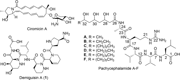
Demiguisin A (1): white solid, UV (acetonitrile), λmax 214 nm; NMR spectral data (DMSO-d6), see Supplementary Table 7 and Supplementary Figs. 7–12; HR-ESI-MS (Supplementary Table 9) m/z 694.3510 [M + H]+ calcd. for C30H47N9O10 at 694.3524 ∆ppm 2.02.
Demiguisin B (2): white solid; UV (acetonitrile), λmax 214 nm; NMR spectral data (DMSO-d6), see Supplementary Table 7 and Supplementary Figs. 13–17; HR-ESI-MS (Supplementary Table 9) m/z 678.3571 [M + H]+ calcd. for C30H47N9O9 at 678.3575, ∆ppm 1.77.
Demiguisin C (3): white solid powder; UV (acetonitrile), λmax 214 nm; NMR spectral data (DMSO-d6), see Supplementary Table 8 and Supplementary Figs. 18–21; HR-ESI-MS (Supplementary Table 9) m/z 637.3299 [M + H]+ calcd. for C28H44N8O9 at 637.3309, ∆ppm 1.57.
Demiguisin D (4): white solid; (acetonitrile), λmax 214 nm; NMR spectral data (DMSO-d6), see Supplementary Table 8 and Supplementary Figs. 22–25; HR-ESI-MS (Supplementary Table 9) m/z 621.3348 [M + H]+ calcd. for C28H45N8O8 at 621.3360, ∆ppm 1.93.
Demiguisin E (5): white solid; UV (acetonitrile), λmax 214 nm; NMR spectral data (DMSO-d6), see Supplementary Table 8 and Supplementary Figs. 26–28; HR-ESI-MS (Supplementary Table 9) m/z 621.3378 [M + H]+ calcd. for C28H45N8O8 at 621.3360, ∆ppm 2.90.
Demiguisin F (6): white solid; UV (acetonitrile), λmax 214 nm; NMR spectral data (DMSO-d6), see Supplementary Table 8 and Supplementary Figs. 29–31; HR-ESI-MS (Supplementary Table 9) m/z 605.3400 [M + H]+ calcd. for C28H45N8O7 at 605.3411, ∆ppm 1.82.
Biological activity of demiguisin derivativesKeratin azure assay
Seed cultures of PS_44_ISF1 and NBC_01488 were grown in 100 mL of PBD for 7 days. After incubation, the cell pellets were washed in 0.9% (w/v) NaCl solution and resuspended in the respective feather medium to an OD600 = 0.7. Thereafter, 5 mL of standardized culture volumes were mixed with either 100 mL of FB or FMB (Supplementary Table 1). Three technical replicates were prepared and placed on a thermoshaker at 180 rpm for 7 days at 30 °C, as well as FB and FMB only for the controls.
Following incubation, the keratinolytic activity was determined by preparing keratin azure (Sigma-Aldrich; cut into small pieces, washed twice with deionized water, and dried). Then, 10 mg of keratin azure was mixed with 800 µl of Tris-HCl (pH 8), followed by the addition of 500 µL of culture supernatant containing extracellular enzymes. The reaction mixtures were incubated for 3 h at 30 °C with shaking at 300 rpm. The reaction was terminated by centrifugation at 10,000 rpm for 10 min at 4 °C. Subsequently, 200 µL of the supernatant from each replicate was transferred into a 96-well plate, and the absorbance was measured at 595 nm. The control FB and FMB were used without additives as the negative control, and 1 µL of protease K was added for the positive control. A calibration curve was created for each medium individually using the following values: 0% for negative control, 100% for positive control. Keratinolytic activity of the samples was subsequently expressed relative to these reference values, allowing comparison of keratin degradation between media and strains32,33.
Activity of fractions against feather degrading bacteria
SPE fractions (10 mg/mL in 50% MeOH, diluted to 1 mg/mL in water) were tested for their activity against Kocuria sp. LS19, Curtobacterium sp. LS33, Curtobacterium sp. LS34, and Kocuria sp. LS38 using a disc diffusion assay34. All test microorganisms were inoculated into Mueller-Hinton broth (MHB) and were incubated at 37 °C for 18–24 h. Following incubation, 100 μL of the bacterial suspension (OD600 = 0.08–0.13) was spread-plated onto Mueller-Hinton agar (MHA, Merck) to create a microbial lawn. On each plate, 6 mm blank discs (Oxoid) were added using a sterile needle, and 50 µL compound solution (50 µg) was applied. A negative control of 10% MeOH was included for each strain, while 50 µL of rifampicin (1 mg/mL) was included as a positive control. Antimicrobial activity was determined by measuring the inhibition zone surrounding the disc in mm.
Bacterial strains and growth conditions for activity of pure compounds
All bacterial isolates were handled following standard microbiological procedures. M. tuberculosis ATCC 25177, M. avium ATCC 700898, S. aureus ATCC 29213 and E. coli ATCC 25922 were purchased from the American Type Culture Collection (ATCC), USA. M. intracellulare ATCC 13950 and M. abscessus ATCC 19977 were acquired from the German Collection of Microorganisms and Cell Cultures (DSMZ). E. coli WO153 strain was generously provided by the Antimicrobial Discovery Center, Department of Biology, Northeastern University. Additional strains were obtained from our internal collection and cultured under conditions recommended by their respective depositors.
Antimicrobial susceptibility testing
The minimum inhibitory concentrations (MICs) of demiguisins were determined using the standard broth microdilution method in Mueller-Hinton Broth II (cation-adjusted; Sigma) according to EUCAST guidelines. Briefly, 75 µL of bacterial suspension (~4 × 10⁵ colony-forming units (CFU)/mL) in Mueller-Hinton Broth II was added to each well of a 96-well plate, along with 75 µL of serially diluted compound solutions (starting concentration 64 µg/mL). Plates were incubated at 37 °C for 18–24 h, and the MIC was reported as the lowest concentration that inhibited visible bacterial growth.
For mycobacterial strains, MIC determination was performed in 7H9 complete medium (BD Difco; Becton Dickinson) supplemented with 10% oleic acid-albumin-dextrose-catalase (OADC; BD), as described previously35. A single-cell suspension of mycobacterial cultures was prepared in PBS and adjusted to a final concentration of 10⁵ CFU/mL for M. tuberculosis and M. marinum and 5 × 10⁴ for M. avium, M. intracellulare, and M. absecuss. Compounds were tested in two-fold serial dilutions with a final DMSO concentration of 1%. Plates were incubated at 37 °C under conditions appropriate for the growth of the respective mycobacterial species, and the MIC was determined as the lowest concentration showing no visible growth.
Cytotoxicity studies using HepG2 cells
HepG2 cells (human hepatoblastoma cell line; DSMZ cell culture no.: ACC 180) were cultured according to the supplier’s instructions in Dulbecco’s modified Eagle medium (DMEM) supplemented with 10% fetal bovine serum. Cytotoxicity of the compounds was assessed as described previously36. Briefly, cells were seeded at 6 × 10³ cells per well in 96-well plates with 120 µL of complete medium. After a 2 h equilibration period, serially diluted compounds were added in 60 µL of complete medium. Compounds, solvent control, and doxorubicin (used as a reference compound) were tested in duplicate in two independent experiments. Following 5 days of incubation, 20 µL of MTT (5 mg/mL thiazolyl blue tetrazolium bromide in PBS) was added to each well, and the plates were incubated for an additional 2 h at 37 °C. The medium was then removed, and the cells were washed with 100 µL PBS. Formazan crystals were dissolved by adding 100 µL of 2-propanol/10 N HCl (250:1) was added. Absorbance was measured at 570 nm using a microplate reader (Tecan Infinite M200Pro). Cell viability was calculated relative to the solvent control, and IC50 values were determined by sigmoidal curve fitting using GraphPad PRISM 8 (GraphPad Software).
BLASTp and multiple protein sequence alignment
To gain better insight into the biosynthetic pathway of a non-proteinogenic amino acid (Piz-1) in the demiguisin derivatives, protein sequences corresponding to DemCYP450 (Accession no. CP109434.1), DmgCYP450 (Accession no. JANUXO000000000.1), PlyM_CYP450 (Accession no. KF386858.1), and HmtN_CYP450 (Accession no. FR823394.1) were retrieved from antiSMASH genome mining results. Pairwise sequence identity between DemCYP450 and the other CYP450 homologues was initially assessed using BLASTp (NCBI) analysis. To further evaluate sequence conservation, the amino acid sequences were aligned using Clustal Omega via the EMBL-EBI web server (https://www.ebi.ac.uk/jdispatcher/msa/clustalo) with default parameters37. The resulting multiple sequence alignment (MSA) was generated in Clustal format and used to assess sequence identity, similarity, and conserved motifs across the CYP450 proteins. The MSA output was subsequently visualized using MView, which produced a formatted alignment figure highlighting conserved residues and regions of divergence. Sequence identity and similarity values were extracted from the Clustal Omega output to support comparative analysis of CYP450 homologues.