
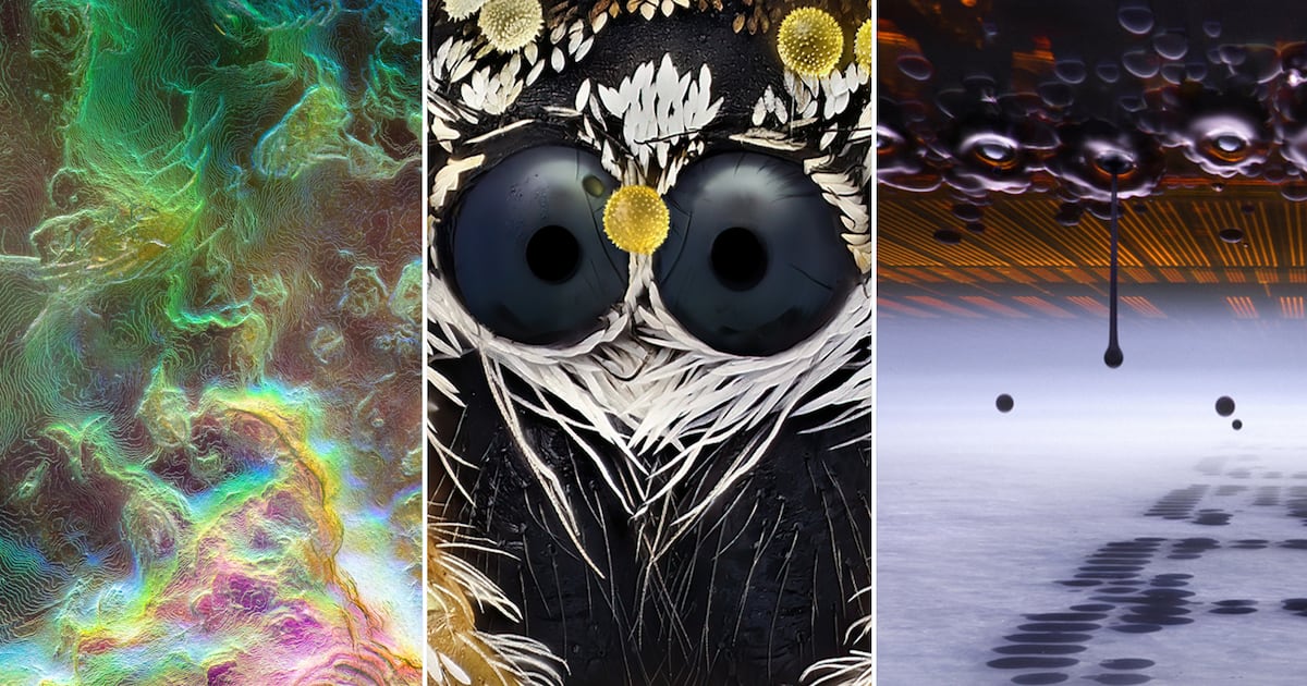
From close-ups of sea snails, mouse eyes and jumping spiders to what ink looks like in your inkjet printer when it’s at work, these are the top 20 best photos of the 2026 Nikon Small World Photomicrography Competition.
Published: October 26, 2026 at 10:39a.m. EDT
 The winners of the 2026 Nikon Small World Photomicrography Competition From close-ups of sea snails to what ink looks like in your inkjet printer when it’s at work, these are the top 20 best photos of the 2026 Nikon Small World Photomicrography Competition.
(Courtesy Nikon Small World)
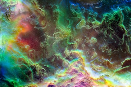 20th Large sea snail (abalone) shell
Photo by:
Justin Theodore Benttinen
Benttinen Photography
Berkeley, California, USA
Techniques
• Image Stacking
• Reflected Light
Magnification
• 20X (Objective Lens Magnification)
(Courtesy Nikon Small World)
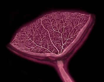 19th Cross-section of a mouse eye showing retinal blood vessels and the optic nerve
Photo by:
Satu Paavonsalo, Dr. Sinem Karaman
University of Helsinki
Individualized Drug Therapy Research Program
Helsinki, Uudenmaan lääni, Finland
Techniques
• Confocal
Magnification
• 20X (Objective Lens Magnification)
(Courtesy Nikon Small World)
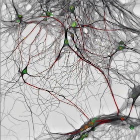 18th 14-day-old mouse hippocampal neurons showing nuclei (green) and synaptic proteins (red)
Photo by:
Bilal Akhtar
Institute of Molecular Biology, Mainz
Department of RNA Biology
Mainz, Germany
Techniques
• Confocal
Magnification
• 40X (Objective Lens Magnification)
(Courtesy Nikon Small World)
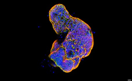 17th Lab-grown human intestinal tissue showing glowing, brick-like epithelial cell connections that form the intestine’s protective barrier
Photo by:
Dr. Gauri Kulkarni
Wake Forest School of Medicine
Wake Forest Institute for Regenerative Medicine (WFIRM)
Winston-Salem, North Carolina, USA
Techniques
• Confocal
Magnification
• 20X (Objective Lens Magnification)
(Courtesy Nikon Small World)
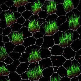 16th Embryonic Xenopus skin cells, showing cilia (green), centrioles (red), and cell membranes (white)
Photo by:
Pierre Nouaux, Laurent Kodjabachian, Camille Boutin
Institut de Biologie du Développement de Marseille (IBDM)
Marseille, France
Techniques
• Confocal
• Fluorescence
Magnification
• 63X (Objective Lens Magnification)
(Courtesy Nikon Small World)
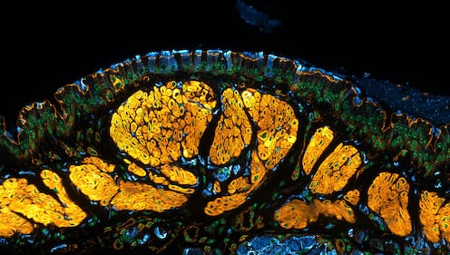 15th Mouse trachea showing tiny hair-like cells that move mucus and nearby nerves (blue) associated with the muscles (yellow)
Photo by:
Dr. Julien Resseguier
University of Oslo
Department of Biosciences
Oslo, Viken, Norway
Techniques
• Confocal
• Deconvolution
• Fluorescence
Magnification
• 60X (Objective Lens Magnification)
(Courtesy Nikon Small World)
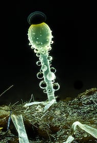 14th Pilobolus fungi on sheep dung
Photo by:
Michael Crutchley
Pembrokeshire, Southwest Wales, United Kingdom
Techniques
• Image Stacking
Magnification
• 20X (Objective Lens Magnification)
(Courtesy Nikon Small World)
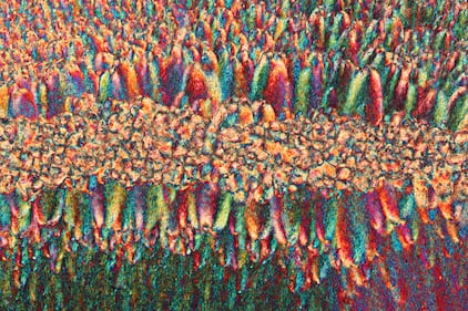 13th Recrystallized estrogen
Photo by:
Sebastian Sparenga
McCrone Research Institute
Chicago, Illinois, USA
Techniques
• Polarized Light
Magnification
• 10X (Objective Lens Magnification)
(Courtesy Nikon Small World)
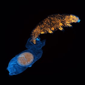 12th A tardigrade molting and laying an egg in the old skin
Photo by:
Dr. Gonzalo Quiroga Artigas
CRBM-CNRS
Montpellier, Hérault, France
Techniques
• Confocal
Magnification
• 40X (Objective Lens Magnification)
(Courtesy Nikon Small World)
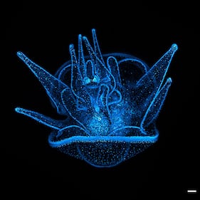 11th 20 day old sea urchin larva with blue fluorescent nuclei
Photo by:
Svenja Carolin Kling
Scripps Institution of Oceanography
Center for Marine Biotechnology and Biomedicine
La Jolla, California, USA
Techniques
• Confocal
Magnification
• 20X (Objective Lens Magnification)
(Courtesy Nikon Small World)
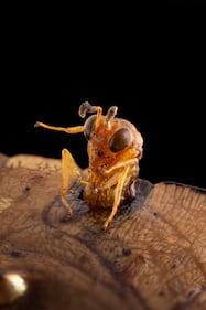 10th A parasitoid wasp (Conura sp.) emerging from a butterfly chrysalis (Opsiphanes sp.)
Photo by:
Michael Parra Puentes
Macrotipos
Bogotá, Cundinamarca, Colombia
Techniques
• Image Stacking
Magnification
• 3.7X (Objective Lens Magnification)
(Courtesy Nikon Small World)
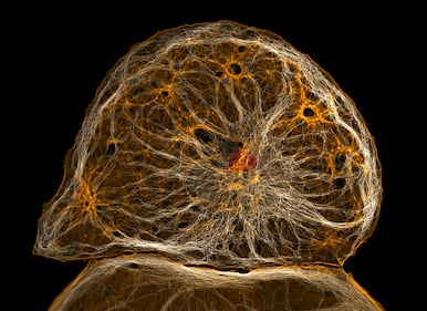 9th Dedifferentiated liver endothelial cell
Photo by:
Dr. Francisco Lázaro-Diéguez
Albert Einstein College of Medicine
Bronx, New York, USA
Techniques
• Confocal
Magnification
• 40X (Objective Lens Magnification)
(Courtesy Nikon Small World)
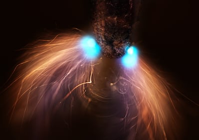 8th Pencil lead during exposure to a high-voltage electrical discharge
Photo by:
Dr. Marcel Clemens
Verona, Veneto, Italy
Techniques
• Image Stacking
Magnification
• 10X (Objective Lens Magnification)
(Courtesy Nikon Small World)
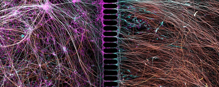 7th Human neurons reprogrammed from skin cells growing in a microfluidic chamber
Photo by:
Dr. Bruno Cisterna, Dr. Eric Vitriol
Medical College of Georgia at Augusta University
Department of Neuroscience & Regenerative Medicine
Augusta, Georgia, USA
Techniques
• Confocal
Magnification
• 20X (Objective Lens Magnification)
(Courtesy Nikon Small World)
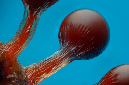 6th Hemitrichia slime mold spore structures (fruiting bodies)
Photo by:
Eric Epstein
Brooklyn, New York, USA
Techniques
• Image Stacking
Magnification
• 10X (Objective Lens Magnification)
(Courtesy Nikon Small World)
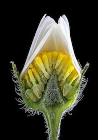 5th Inner structure of a closed daisy
Photo by:
Michael Crutchley
Pembrokeshire, Southwest Wales, United Kingdom
Techniques
• Darkfield
• Image Stacking
Magnification
• 10X (Objective Lens Magnification)
(Courtesy Nikon Small World)
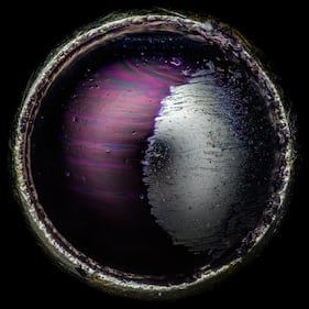 4th Tip of a ballpoint pen
Photo by:
Justin Theodore Benttinen
Benttinen Photography
Berkeley, California, USA
Techniques
• Image Stacking
• Reflected Light
Magnification
• 20X (Objective Lens Magnification)
(Courtesy Nikon Small World)
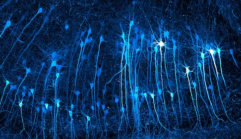 3rd Neurons in an expanded mouse brain
Photo by:
Kaelin Wulf, Rachel Flannery
Allen Institute
Department of Neural Dynamics
Seattle, Washington, USA
Techniques
• Expansion Microscopy
• Fluorescence
• Light Sheet
Magnification
• 5X (Objective Lens Magnification)
(Courtesy Nikon Small World)
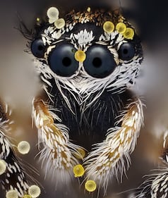 2nd Jumping spider (Salticus scenicus) with pollen
Photo by:
John-Oliver Dum
Medienbunker Produktion
Bendorf, Rheinland Pfalz, Germany
Techniques
• Image Stacking
Magnification
• 20X (Objective Lens Magnification)
(Courtesy Nikon Small World)
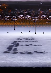 1st Microscopic view of an inkjet printer depositing ink onto paper
Photo by:
Ole Bielfeldt
Macrofying
Cologne, North Rhine-Westphalia, Germany
Techniques
• Slow-Motion
• Image Stacking
Magnification
• 20X (Objective Lens Magnification)
(Courtesy Nikon Small World)