
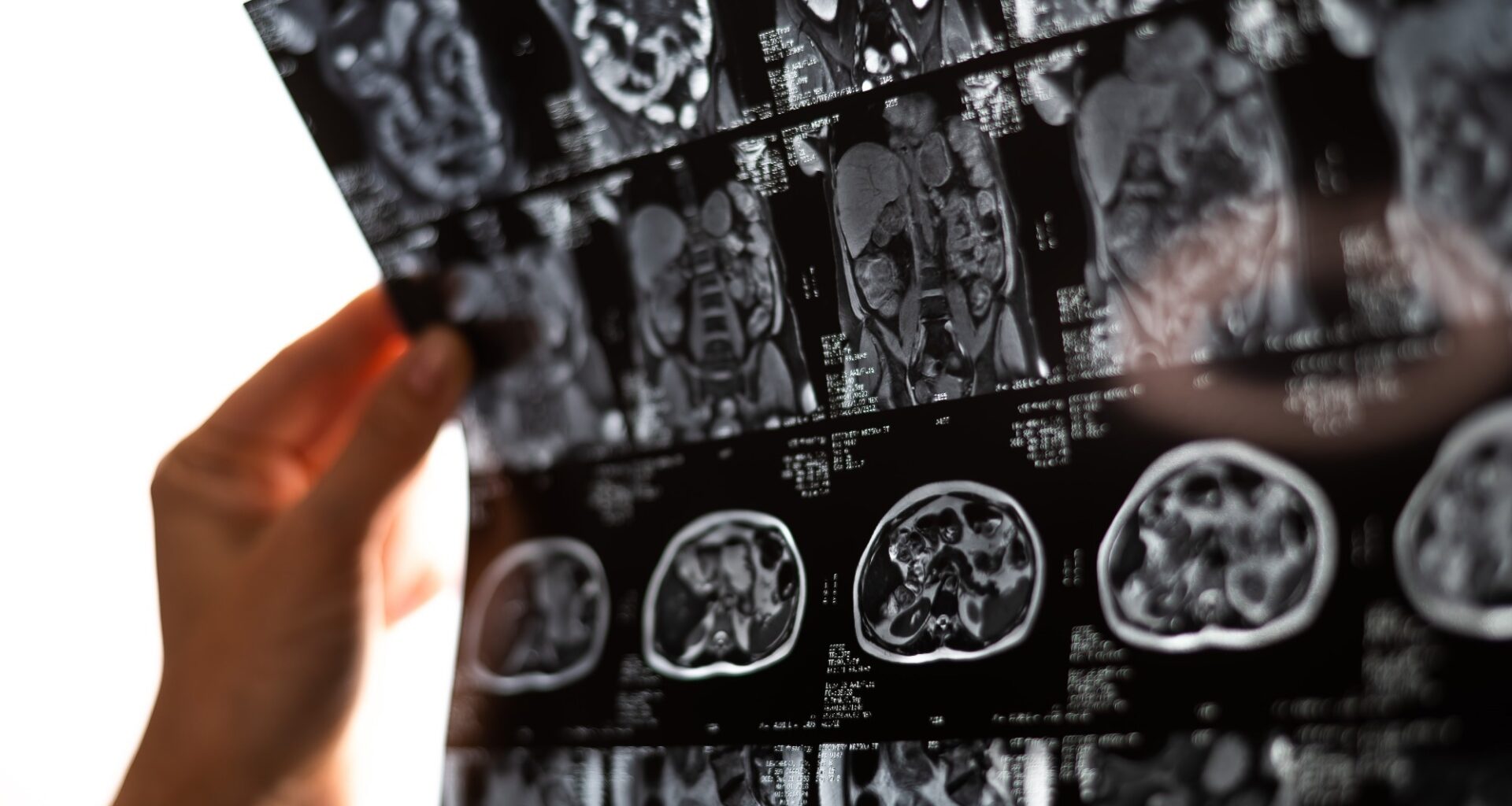
Analyzing heart scans in seconds rather than minutes, AI slightly outperformed expert clinicians in predicting mortality across more than 27,000 patients, but researchers caution that its clinical benefits still need prospective validation.
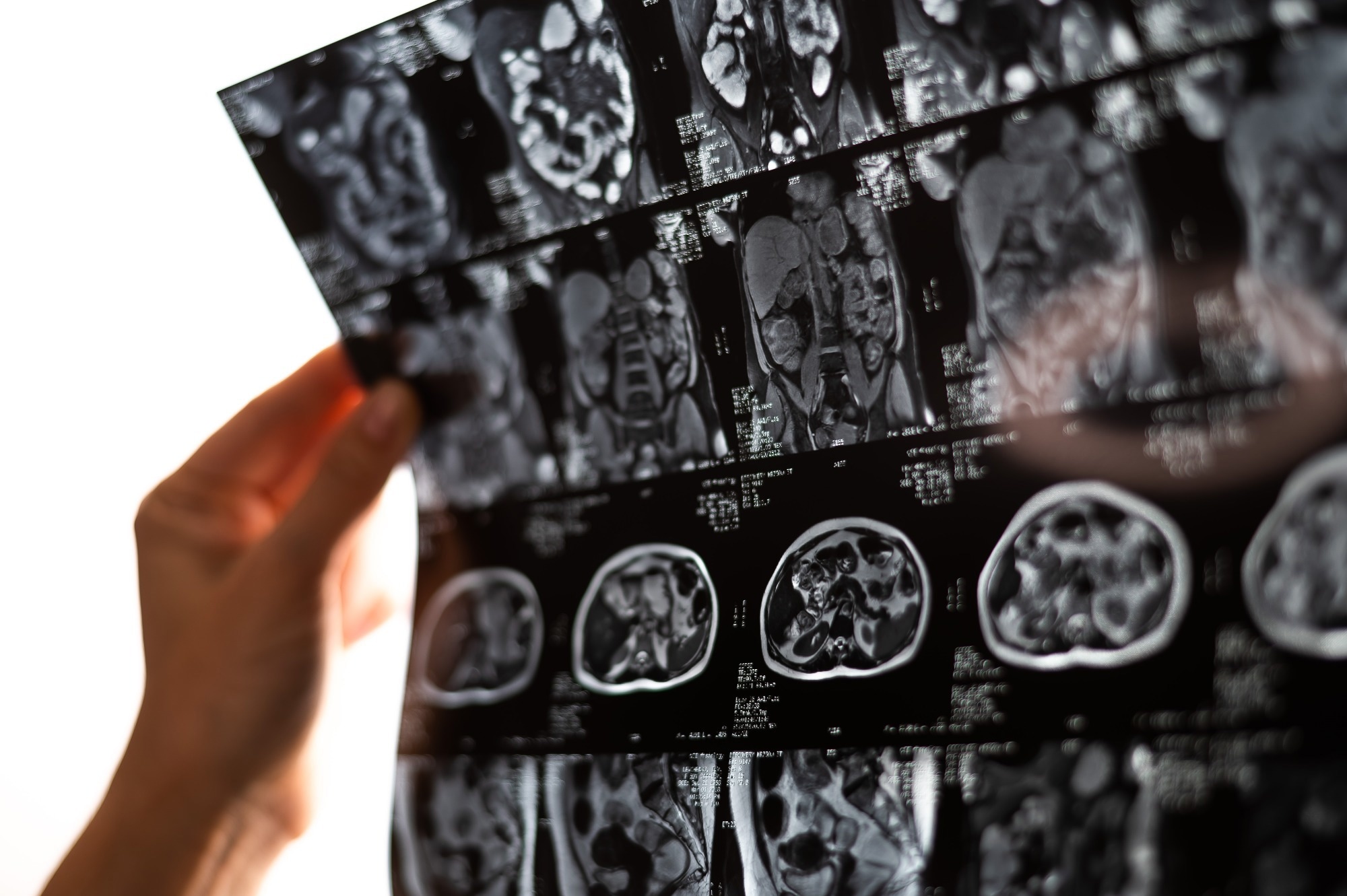 Study: Performance of artificial intelligence versus clinician left-ventricular segmentation of cardiac MRI for mortality prediction: a retrospective, multicentre, prognostic study. Image credit: Reshetnikov_art/Shutterstock.com
Artificial intelligence (AI)-derived measurements of left-ventricular ejection fraction from cardiac MRI scans predicted all-cause mortality slightly better than clinician-derived measurements, according to a retrospective study published in The Lancet Digital Health. The findings suggest that AI could help streamline cardiac function assessments, although its clinical benefits require further investigation.
Can AI improve how cardiac MRI predicts mortality?
Left-ventricular ejection fraction is a key measure of cardiac function, widely used in clinical decision-making and risk stratification. Cardiac MRI is considered the gold standard for assessing cardiac function, but manual image segmentation by clinicians is subject to observer variability, which may limit measurement precision.
Previous research suggests that AI can reduce measurement variability, improve precision, and substantially shorten image-processing times. However, whether these advantages translate into better prediction of clinical outcomes remains incompletely understood.
The current study investigated whether AI-derived measurements of ejection fraction and other left-ventricular parameters from cardiac MRI scans outperform expert clinician measurements in predicting all-cause mortality.
AI predicts mortality marginally better than expert clinicians
The retrospective study included 27,382 patients who underwent cardiac MRI at two centers in the UK and USA. During a median follow-up of 5.6 years, 13% died.
AI-derived ejection fraction showed better statistical model fit and slightly better discrimination for all-cause mortality than clinician-derived measurements.
Before adjusting for age and sex, the AI-based model achieved a C-index of 0.669, compared with 0.637 for clinician measurements. After adjustment, the difference narrowed substantially, with C-index values of 0.781 and 0.777, respectively. Although statistically significant, the improvement in mortality discrimination was small.
Adding AI-derived ejection fraction to a model containing clinician measurements substantially improved model fit. Conversely, adding clinician measurements to an AI-based model produced only a slight improvement. These findings suggest that AI measurements captured most, but not all, of the prognostic information provided by clinician measurements.
The AI algorithm also processed cardiac MRI scans considerably faster, requiring approximately 3–4 seconds per patient. In comparison, clinician segmentation took a median of 13 minutes per patient in an earlier study.
However, AI measurements were not error-free. In a randomly selected sample of 600 patients, segmentation errors occurred in six cases, corresponding to a 1% error rate. Errors were considerably more frequent among scans with extreme ejection fraction measurements, particularly very low values, highlighting the importance of clinician oversight.
AI also produced systematically different measurements from clinicians, with a lower median ejection fraction of 57.8%, compared with 62.1%. However, the study did not establish which measurements were more anatomically accurate against an independent reference standard.
Small predictive gains highlight promise and limitations of AI
The findings highlight AI’s potential to accelerate cardiac MRI analysis, reduce clinician workload, and modestly improve mortality risk prediction.
Even small improvements in prognostic performance could potentially be valuable when applied across large patient populations. However, the clinical importance of the observed differences remains uncertain.
The researchers also examined predictive performance across different cardiac conditions.
In patients with obstructive coronary artery disease, AI-derived ejection fraction showed better mortality model fit than clinician measurements. Adding AI measurements to clinician-derived measurements further improved the model, whereas adding clinician measurements to AI-derived measurements did not.
In patients with non-ischemic cardiomyopathy, AI showed a smaller advantage in model fit, but adding AI-derived ejection fraction to clinician measurements did not significantly improve prediction.
In contrast, clinician-derived ejection fraction performed slightly better in patients with hypertrophic cardiomyopathy. The researchers suggested that the AI algorithm’s segmentation approach might not fully capture certain anatomical features, including hypertrabeculation and enlarged papillary muscles.
However, left-ventricular volume measurements were generally poor predictors of mortality in this subgroup, regardless of whether AI or clinicians obtained them.
The overall findings are consistent with previous research reporting better mortality prediction using AI-derived ejection fraction from echocardiography in patients hospitalized with acute COVID-19.
The study also highlights the potential of large-scale clinical datasets for evaluating AI-based prognostic tools under real-world imaging conditions. Nevertheless, several limitations warrant consideration.
The analysis was retrospective and did not include cause-specific mortality data. Differences in clinician segmentation techniques could also have contributed to the observed advantage of AI. Furthermore, the researchers evaluated statistical model fit and mortality discrimination but did not assess risk calibration or whether AI-guided measurements improve clinical decisions or patient outcomes.
Although the study included patients from both the UK and the USA, most participants came from one UK center, and the authors did not evaluate performance across ethnic groups. Several authors also disclosed financial relationships with MycardiumAI, the company associated with the segmentation technology used in the study.
The researchers concluded that AI-derived ejection fraction could be useful as a first-line measurement approach with clinician oversight. However, prospective studies are needed to establish its clinical utility, cost-effectiveness, and impact on patient care.