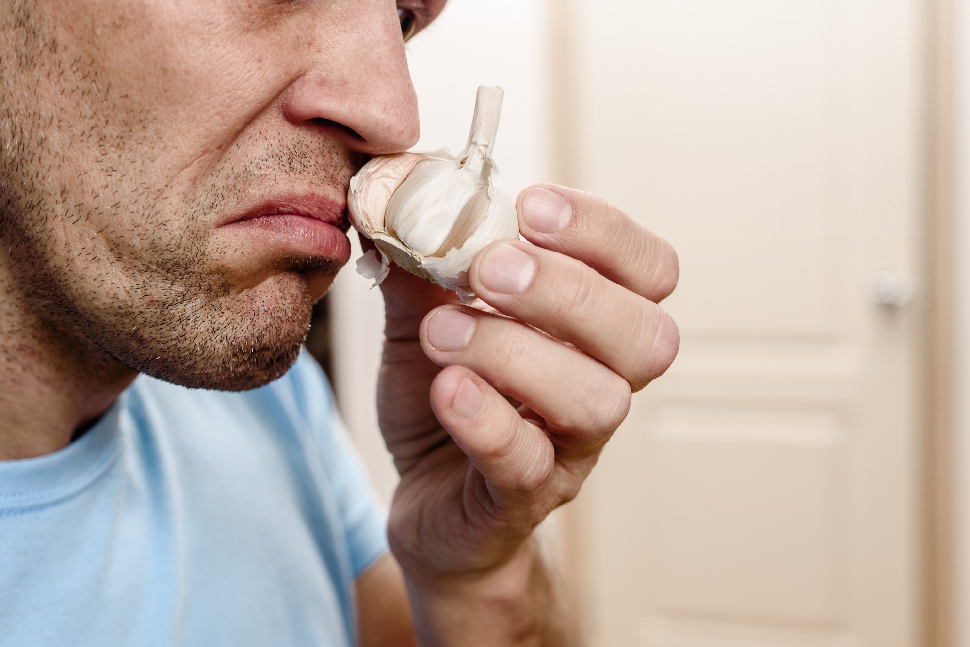
MRI scans reveal that people who lose their sense of smell after COVID-19 show measurable changes in brain regions tied to emotion and sensory processing, offering new clues to the lingering effects of long COVID.
 Study: Alterations of the amygdala in post-COVID olfactory dysfunction. Image credit: Denys Kurbatov/Shutterstock.com
In a recent study published in Scientific Reports, researchers investigated the structural integrity and connectivity of white matter pathways in olfactory-related brain regions in patients with post-coronavirus disease 2019 (COVID-19) olfactory dysfunction (OD).
The olfactory aspect of COVID-19 has attracted considerable research attention. While many people’s acute COVID-19 symptoms subside after a few weeks, a substantial proportion struggle with persistent health issues, including persistent OD, long after the infection has cleared. Understanding the factors underlying this phenomenon is crucial to developing effective management strategies and supporting those affected.
Previous studies have reported heterogeneous findings across patient populations, and this new work specifically focused on individuals with mild, non-hospitalized COVID-19 cases to avoid confounding factors related to severe disease or treatment effects.
About the study
In the present study, researchers investigated the structural integrity and connectivity of white matter pathways in brain regions related to olfactory processing in patients with post-COVID OD. Sixty-one individuals from the COVIDOM study, which investigated COVID-19 long-term effects, were included. All participants had a confirmed SARS-CoV-2 infection at least six months ago.
Individuals with pregnancy, medical implants incompatible with magnetic resonance imaging (MRI), or claustrophobia were excluded. The Sniffin’ Sticks test was used to assess olfactory function, and the threshold-discrimination-identification (TDI) score was calculated from its three subtests: threshold, discrimination, and identification. Participants with persistent OD after SARS-CoV-2 infection, defined as a TDI score < 31, were included in the post-COVID OD (PC-OlfDys) group.
After their SARS-CoV-2 infection, individuals with normosmia were included as post-COVID normosmic (PC-N) controls. Participants completed questionnaires covering demographic data, medical history, the course of acute SARS-CoV-2 infection, and smoking status. Participants also rated their sense of smell on a 10-point scale before, during, and after their SARS-CoV-2 infection.
The generalized anxiety disorder 7 (GAD-7) questionnaire and patient health questionnaire-depression (PHQ-8) were also completed. Cognitive impairment was assessed using the Montreal Cognitive Assessment test (MoCA). Diffusion tensor imaging, an advanced MRI technique, was performed, and tract-based spatial statistics (TBSS) were used for voxelwise analyses of fractional anisotropy (FA), mean diffusivity (MD), radial diffusivity (RD), and axial diffusivity (AD) data.
Further, region-of-interest (ROI)-based analyses were conducted. The Mann-Whitney U-test was used for intergroup comparisons. An independent samples t-test compared FA, RD, AD, and MD values from the ROI between groups. A bivariate Pearson’s correlation analysis was performed between the questionnaire and diffusion values.
The whole-brain TBSS analyses did not show any significant differences between groups after correction for multiple comparisons, so the ROI-based approach was key to identifying subtle, region-specific changes.
Findings
The PC-OlfDys and PC-N groups included 31 and 30 participants, aged 44 and 44.4 years, on average, respectively. The sense of smell was rated significantly lower by PC-OlfDys subjects during and after SARS-CoV-2 infection compared to PC-N controls. Further, 38% of PC-OlfDys subjects reported perceiving odors differently than before, indicating parosmia (distorted odor perception), while none in the control group reported changes in smell perception.
In the Sniffin’ Sticks test, there were significant differences between the two groups for the TDI score, albeit controls performed better. The average PHQ-8 score was nine in the PC-OlfDys group and two in the PC-N group. The average GAD-7 score was six for the PC-OlfDys group and one for controls. There were no significant differences in MoCA between the two groups.
Further, while whole-brain TBSS showed no significant group differences, ROI-based analyses revealed that left amygdala FA values were significantly higher in the PC-OlfDys group than in controls. Moreover, the PC-OlfDys group had higher right amygdala RD values than controls. Other ROIs showed no significant differences in diffusion values.
There was no correlation between the overall TDI score and any diffusion value in the PC-OlfDys group. However, identification, discrimination, and threshold (subtest) scores were negatively correlated with AD, MD, and RD values in the anterior piriform cortex, left amygdala, and right putamen, respectively, in the PC-OlfDys group. In controls, the TDI score showed a positive correlation with right putamen MD values, and the identification score showed a negative correlation with left amygdala RD values.
Further, in the PC-OlfDys group, OD duration was positively correlated with AD values in the left amygdala and MD values in the left posterior piriform cortex and left amygdala. The PHQ-8 score was negatively correlated with left putamen AD values and left amygdala FA values in the PC-OlfDys group. The GAD-7 score was negatively correlated with left amygdala FA values in PC-OlfDys subjects. PHQ-8 and GAD-7 scores showed no correlations in controls.
Conclusions
The findings indicate significant changes in the brain associated with persistent OD after SARS-CoV-2 infection. Alterations were identified in several olfactory brain regions, especially in the amygdala, piriform cortex, and putamen, where increased FA values may reflect enhanced myelination and reorganization of white matter pathways. In contrast, elevated RD values could indicate microstructural alterations or disrupted myelin integrity.
DTI findings should not be interpreted as direct evidence of neurodegeneration but rather as possible indicators of adaptive or compensatory processes within olfactory circuits.
Moreover, the longer the OD post-infection, the greater the DTI changes in critical olfactory circuits. These neural alterations were also linked to higher anxiety and depression scores, suggesting a potential bidirectional relationship between persistent olfactory loss and emotional well-being, though causality remains unclear.

