
“We would like to more fully understand how these patterns are disrupted during disease, for hearing research on noise damage and aging,” said Manor. “By visual inspection we can see that the normal bundle patterns tend to fall apart. Some of them become longer and others shorter. We want to understand exactly how this is happening.”
VASCilia provides a method of visualizing and quantifying these cells in 3D using AI advancements. Kassim, a computer scientist and a Schmidt AI Postdoctoral Fellow, trained VASCilia on sets of stereocilia data obtained through expert-annotated datasets from mice. Five deep learning-based models streamlined the tool’s cell analysis process.
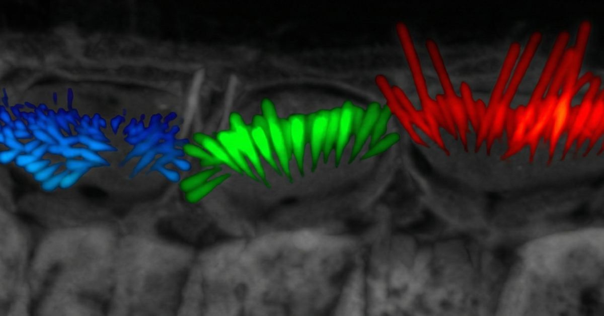
“We’ve reduced the amount of time it takes to analyze the length of these cells by a factor of 50, enabling many additional 2D and 3D quantitative measurements that can be acquired in minutes — work that would otherwise require years of manual analysis,” said Kassim. “VASCilia can also generate other perspectives, such as the orientation of the cells, which is useful since hair bundles sometimes don’t align after aging or damage. Further, VASCilia can detect and quantify subtle patterns of cellular disorganization that are difficult for humans to measure manually.”
The researchers hope the open-source nature of VASCilia will eventually lead to a comprehensive atlas of cochlea hair cell images.
“Ultimately, this initiative will support the development of foundational models adaptable to various species, markers and imaging scales to accelerate advances within the hearing research community,” the authors conclude in the paper.
The authors of the study are: Yasmin M. Kassim, David B. Rosenberg, Samprita Das, Xiaobo Wang, Zhuoling Huang, Samia Rahman, Ibraheem M. Al Shammaa, Samer Salim, Kevin Huang, Alma Renero, Yuzuru Ninoyu, Rick A. Friedman, Artur Indzhykulian and Uri Manor.
The research was supported by the Chan Zuckerberg Initiative DAF (CZI Imaging Scientist Award DOI:10.37921/694870itnyzk), the National Science Foundation (NSF NeuroNex Award 2014862), the David F. And Margaret T. Grohne Family Foundation, the G. Harold & Leila Y. Mathers Foundation and the National Institute on Deafness and Other Communication Disorders (NIDCD grants R018566-03S1 and R01 DC021075-01). Microscopy was supported by the Waitt Advanced Biophotonics Core of the Salk Institute with funding from the Waitt Foundation, the NIH National Cancer Institute (NCI CCSG P30 014195) and the San Diego Nathan Shock Center.
Learn more about research and education at UC San Diego in: