
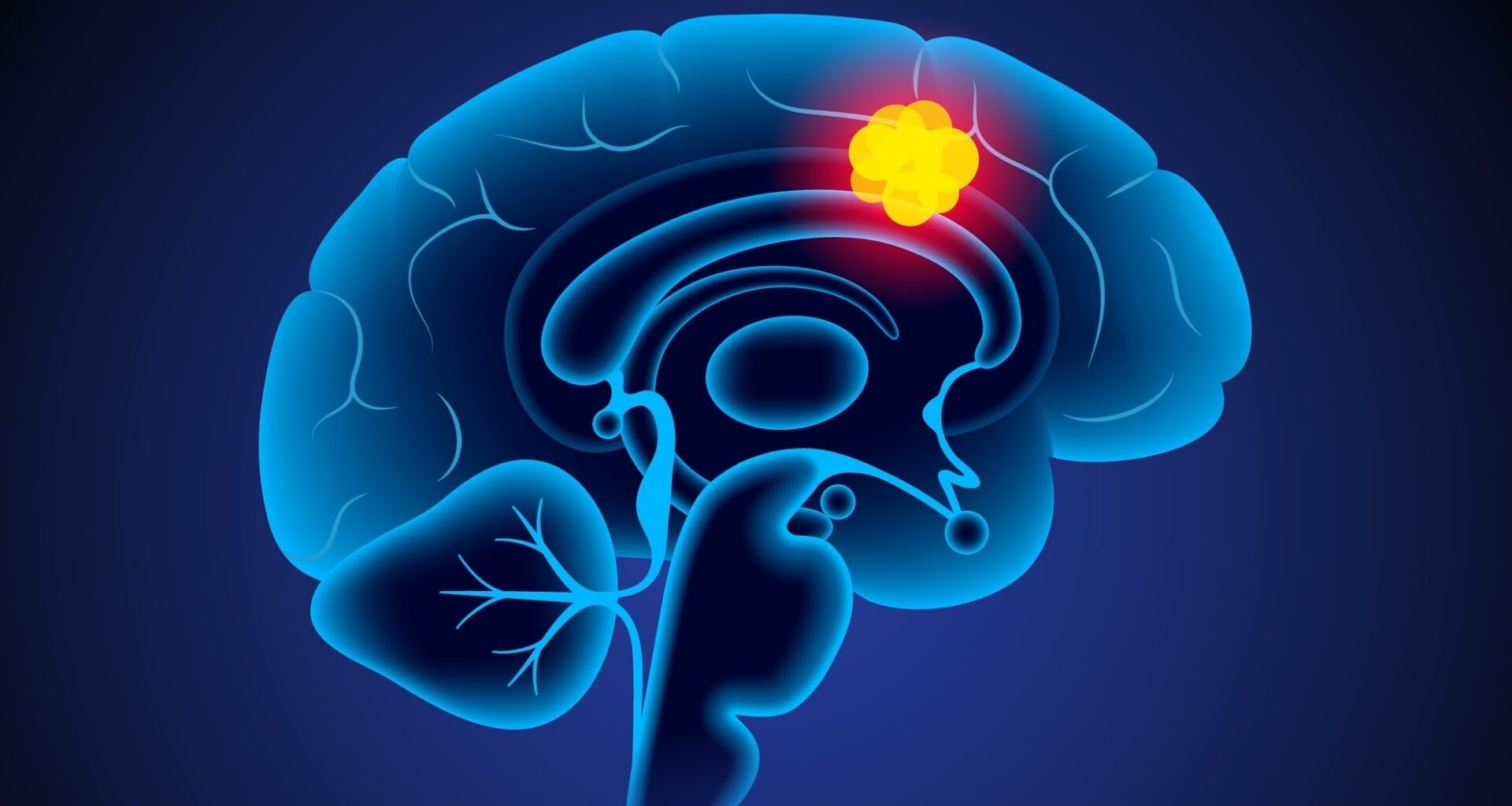
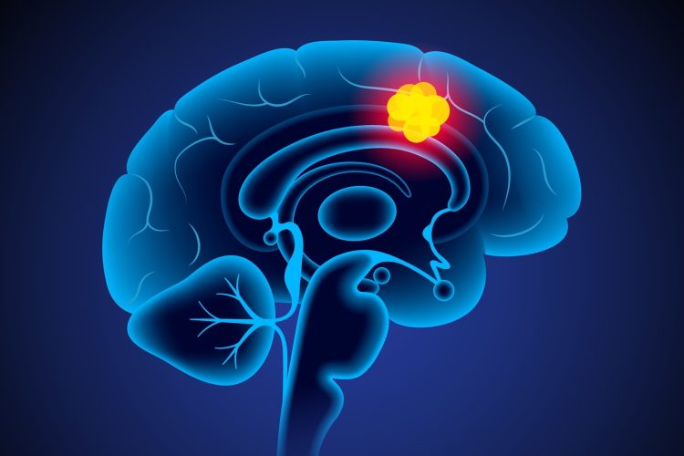
Researchers at the University of Cincinnati are developing an immune-stimulating wafer to be used after brain tumour surgery, aiming to overcome long-standing barriers in the treatment of glioblastoma and inform new personalised therapies.
A multidisciplinary team at the University of Cincinnati Cancer Center has secured a $40,000 Ride Cincinnati grant to develop a novel approach aimed at improving outcomes for patients with glioblastoma. The project will explore a delayed release wafer designed to stimulate the central nervous system immune response following surgery.
Tackling a lethal brain cancer
Glioblastoma is one of the most challenging cancers to treat, with only 5-7 percent of patients surviving five years after diagnosis.
Researchers have long struggled to develop effective therapies because of two major barriers. The blood brain barrier protects the brain from infection but also blocks many cancer drugs from reaching tumour cells. In addition, the central nervous system has what scientists describe as a ‘cold’ immune microenvironment, meaning it is difficult to trigger an immune response capable of eliminating cancer cells left behind after surgery.
Glioblastoma is one of the most challenging cancers to treat, only 5 percent to 7 percent of patients survive five years after diagnosis.
Current surgical options include implantable wafers that deliver radiation or non-specific cell-killing agents. However, these approaches are costly and have shown limited benefit for patients.
“After surgery to remove the tumour, we have unencumbered access to a resection cavity that we know microscopically is invaded by tumour cells,” said Jonathon Forbes, MD, Associate Professor and Residency Programme Director in the Department of Neurosurgery in UC’s College of Medicine and a UC Gardner Neuroscience Institute neurosurgeon. “Why not use this access to enhance the central nervous system’s ability to clear residual tumour cells?”
Activating the immune system
Medical student Beatrice Zucca explained that the first phase of the research focused on identifying a molecule capable of safely activating the brain’s immune defences. The team selected Interleukin-15, known as IL-15.
“IL-15 is exceptionally effective at activating immune populations that are critical for recognising and killing cancer cells,” said Zucca, who worked as a neurooncology research fellow under Forbes’ mentorship. “It improves their survival, expands their numbers and enhances their cell-killing function, making it an ideal candidate for driving a coordinated immune attack against a highly resistant cancer like glioblastoma.”
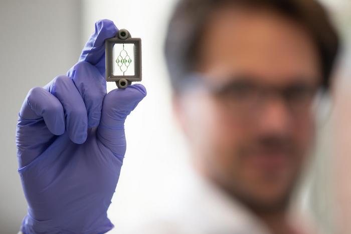
Barrile’s lab designed the first glioblastoma-on-a-chip model that uses bioprinting and 3D printing to integrate human brain cells with glioblastoma cells. Credit: Photo/Andrew Higley/UC Marketing + Brand.
Testing tumours on a chip
Grant funding will enable the team to test the immune-stimulating wafer using glioblastoma-on-a-chip technology developed with Dr Ricardo Barrile.
“An organ-on-a-chip is a miniaturised model of a living organ engineered to incorporate the minimal biological elements needed to recreate specific disease conditions,” said Barrile, Assistant Professor of Biomedical Engineering in UC’s College of Engineering and Applied Science. “Instead of testing drugs on flat plastic dishes or relying solely on animal models – which often fail to predict human results due to genetic disparities – we use 3D bioprinting and microfluidics to build a living model of a human organ.”
An organ-on-a-chip is a miniaturised model of a living organ engineered to incorporate the minimal biological elements needed to recreate specific disease conditions.
Barrile’s laboratory was the first to integrate human brain cells and glioblastoma cells using combined 3D printing and bioprinting. The model includes channels that mimic blood vessels and the immune system, creating what Barrile describes as a more accurate human platform.
“This provides a ‘human-relevant’ platform to test therapies safely and accurately before they reach a patient,” Barrile said. “Integrating the immune system was the missing piece and is the key to capture the natural composition of glioblastoma, which in a patient is typically made up to 30 percent of immune cells. These cells are typically lost during in vitro cell culture.”
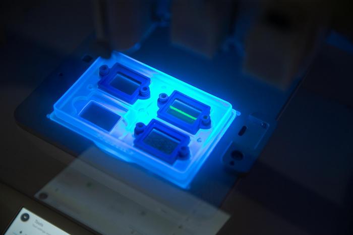
UC biomedical engineer Riccardo Barrile and his students have developed a 3D-printed blood vessel to test new drugs for brain tumours. Credit: Photo/Andrew Higley/UC Marketing + Brand.
Toward personalised treatment
Beyond this study, the technology could support personalised medicine by predicting how individual patients respond to immunotherapy.
“We are building a platform that could eventually predict a specific patient’s response to immunotherapy,” Barrile said. “By using a patient’s own cells on our chip, we can identify the best therapeutic approach for that specific individual before treatment even begins. We are essentially moving from a one-size-fits-all approach to a tailored-to-you strategy.”
Forbes added that parallel research at the UC Brain Tumor Center is exploring focused ultrasound techniques to temporarily open the blood brain barrier.
“It’s very exciting that we’re actually working on both fronts at the University of Cincinnati, trying to find better treatments for glioblastoma,” he said.
For Zucca, the project carries personal significance as well as scientific promise for the future.
“It brings together molecular immunology, biomedical engineering and clinical neurooncology in a way that has profoundly influenced my development as a researcher,” she said. “Most importantly, it represents a tangible step toward therapies that leverage the patient’s own immune system to combat one of the most aggressive cancers known.”
Related topics
Bioengineering, Bioprinting, Cancer research, Central Nervous System (CNS), Cytokines, Drug Delivery, Microfluidic Technology, Oncology, Organ-on-a-Chip, Personalised Medicine, Translational Science