
The skin covering human bodies looks smooth from the outside, but under a microscope, it is shaped by tiny folds called rete ridges.
Scientists have long assumed these structures formed before birth and remained mostly unchanged afterward. New research now shows that assumption was wrong.

These microscopic rete ridges develop shortly after birth, revealing an unexpected early-life window when the skin’s internal architecture is still being built.
The finding reshapes how scientists think about aging, scarring, and skin repair, suggesting that the same biological signals that create these folds early in life could one day help restore them later.
Lifespan of rete ridges
The skin of some mammals retains its rete ridges well into adulthood, revealing a developmental window that had gone undocumented, until now.
By tracking skin architecture across species, work centered at Washington State University (WSU) allowed Ryan Driskell and colleagues to directly observe when the folds first appeared.
Instead of arising during fetal growth, the structures emerged shortly after birth, overturning long-held assumptions about when skin establishes its internal grip.
That narrow window helps explain why the process went unseen for so long and only became clear through closer comparison.
Flattening rete ridges
Rete ridges act like small anchors that help hold the layers of your skin together.
They increase the contact between the outer layer, called the epidermis, and the deeper dermis layer. Fibers, blood vessels, and nutrients fill the tougher dermis and support the surface.
Because these folds create extra grip between the layers, skin can better handle the stretching, pulling, and rubbing it experiences every day.
When rete ridges flatten – something that often happens with aging – the layers do not lock together as tightly. As a result, the skin becomes thinner, sags more easily, and is more prone to bruises and tears.
Finding better skin models
Skin research has long relied on mice and monkeys, but scientists now realize those models missed a key feature.
Much of their trunk skin lacks rete ridges, meaning the epidermis meets the dermis along a flatter boundary that changes how tension and nutrients move.
WSU researchers found that several thick-skinned mammals share the human-style folded structure, while many primate models retain the smoother base. This mismatch likely slowed progress and pointed the field toward better stand-ins.
Because late-stage human fetal samples are rare, pigs became the clearest way to watch when the folds actually form. Partnering with local farmers, the team collected pig skin across development and tracked the first appearance of ridges.
“We expected this structure to be established before birth, so seeing it emerge afterward was a surprise,” said Driskell.
The revised timeline changes how scientists understand when skin architecture develops and suggests that the process may remain more flexible later in life than previously believed.
Signals behind the folds
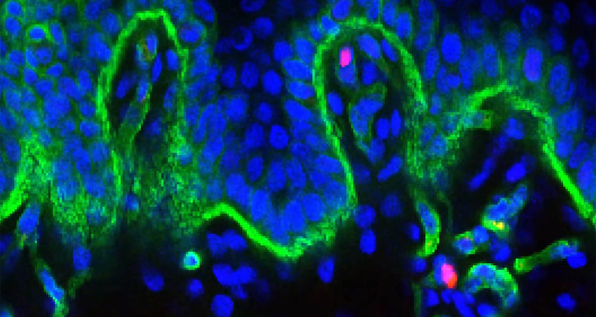
One message rose as ridges formed, and it came from BMP signaling, a bone morphogenetic protein cue that guides cells. BMP activity told skin cells to communicate across layers, pushing them to dip down in connected lines.
Gene activity maps showed that BMP was turned on broadly in ridge-building skin, and mouse finger pads needed it to form folds.
Because ridge networks fade over time, researchers now see BMP as a promising handle for rebuilding strength.
Regrowing ridges after injury
Newborn pig skin carried a striking ability that adult scars rarely show, and rete ridges returned after wounds. Researchers made deep wounds about 1 inch by 1 inch, then watched healing rebuild the folded pattern.
The repaired skin also recreated dermal pockets, small spaces under ridges with extra blood vessels, matching uninjured tissue. That result hints skin can rebuild its deep attachment, but only if the right signals arrive early enough.
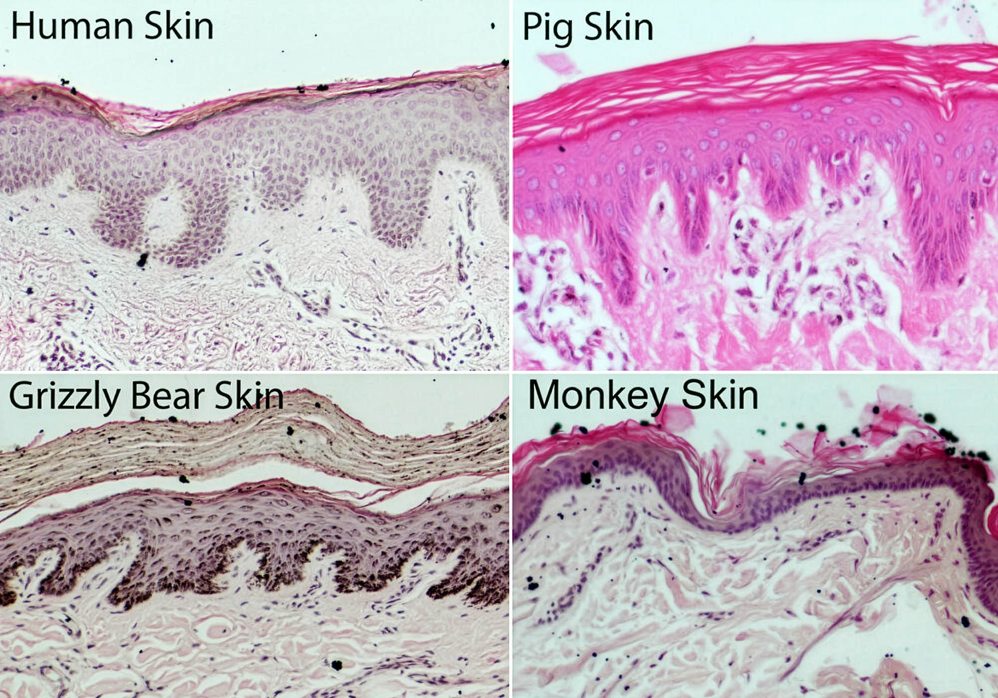 These microscopic views of layers of skin shows how humans, grizzly bears, and pigs (but not monkeys) all share valley-like skin microstructures called rete ridges, which are visible in the bottom sections of these slide images. Credit: Ryan Driskell and Sean Thompson, Washington State University College of Veterinary Medicine. Click image to enlarge.From biology to treatment
Turning ridge formation into a therapy will demand careful control, because growth signals can cause trouble in new places.
The Food and Drug Administration (FDA) has already cleared a BMP-based bone graft, and the approval shows real-world medical use.
“These structures degrade as we age; now we know how they form and have a blueprint to guide future work on restoring them,” said Driskell.
Any skin use would face FDA review again, since extra BMP could trigger swelling or unwanted bone growth.
Aging hits the boundary first
As skin ages, the once-wavy boundary between the epidermis and the dermis gradually flattens, reducing the contact area that helps the two layers stay firmly connected.
Researchers believe this happens partly because cells in the older epidermis divide more slowly, leaving less new tissue available to maintain the deep folds that normally reinforce the junction.
With fewer of these anchoring ridges, the skin’s surface slides more easily over the layer beneath it, making everyday bumps and scrapes more likely to cause bruises or tears.
Scientists are now exploring whether signals such as BMP could safely rebuild these ridges, which might help restore some strength to aging skin and allow wounds to heal with less stress.
Rete ridges and human skin aging
Across mammals, rete ridges appeared in thick-skinned species such as grizzly bears and dolphins, as well as in humans.
Researchers linked these ridge networks to a thicker epidermis, suggesting the folds help exposed skin remain durable when hair cover decreases.
Even large, furry grizzlies retained ridges in wide spaces between hair follicles, showing that hair alone does not erase the pattern.
These comparisons give skin researchers a clearer rulebook for selecting experimental models and identifying which skin traits tend to develop together.
The same research also connected an early-life construction window to a single biological signal and showed that the ridge pattern can return after injury.
Researchers will next test how well they can deliver this signal in adult human skin. After that, regulators will review the results before any potential scar-reduction or anti-aging therapies reach clinical use.
The study is published in Nature.
—–
Like what you read? Subscribe to our newsletter for engaging articles, exclusive content, and the latest updates.
Check us out on EarthSnap, a free app brought to you by Eric Ralls and Earth.com.
—–