
What happens when two critical proteins collaborate during neurodevelopment? A new Molecular & Cellular Proteomics study reveals insights that could transform how we understand childhood brain disorders.
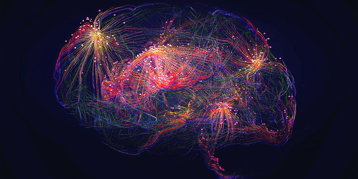
The brain’s intricate structure depends on precisely timed signals that guide cells to differentiate and migrate. When these signals go awry, the consequences can range from developmental disorders to tumor formation. Cortical development, which supports thinking, perception, language, movement and memory, is especially sensitive to these signals.
Wnt signaling and its downstream effector, CTNNB1, play a central role in this process, controlling the growth, development and migration of brain cells. Yet, when Wnt signaling becomes overactive, it can disrupt normal cortical development and, in some cases, lead to brain tumors in children. The study, headed by Jelena Navolić and Julia Neumann at University Medical Center Hamburg-Eppendorf in Germany, uncovered molecular mechanisms underlying specific brain malformations and tumors. The findings also point to potential therapeutic targets.
Building on this framework, the study centers on LIN28A, an oncoprotein, and CTNNB1, which encodes β-catenin, a core component of Wnt signaling. Previous work suggests that LIN28A and Wnt signaling have interconnected roles in brain development, but their precise functional link remained unclear.
To address this gap, Navolić and colleagues investigated how coactivation of LIN28A and Wnt signaling affects cortical development, analyzing structural changes in the cerebral cortex of the mouse. They induced overexpression of Lin28A and stabilized the Ctnnb1 gene in the same population of neural precursor cells, causing accumulation of both LIN28A and CTNNB1 pathways.
The team used nanosecond infrared laser technology to precisely sample tiny regions of mouse brain cortices, allowing them to map protein abundance at unprecedented resolution. They found that in the brain, during embryonic development, disrupted cortical layering and impaired neuron migration.
They also observed changes in the distribution of extracellular matrix, or ECM, receptors RPSA and ITGB1, along with reduced glycosylation of α-dystroglycan. ECM receptors anchor cells to their surroundings, while glycosylation allows α-dystroglycan to bind effectively to ECM proteins.
These alterations weaken cell attachment and signaling, impairing neuron migration — an essential process for neurodevelopment and tissue repair. The defects resemble cobblestone lissencephaly type 2, a rare disorder marked by a bumpy brain surface and neurological deficits.
Overall, the study reveals a previously unrecognized role for LIN28A in maintaining the extracellular matrix during brain development, especially when combined with CTNNB1. Furthermore, the findings suggest that interactions between oncogenes can contribute to both tumor formation and developmental brain disorders. The work also highlights the power of nano-volume spatial proteomics to map protein distributions across tiny brain regions in remarkable detail. These insights may guide future research and therapeutic strategies for rare brain disorders and aggressive pediatric tumors linked to LIN28A and Wnt signaling.