
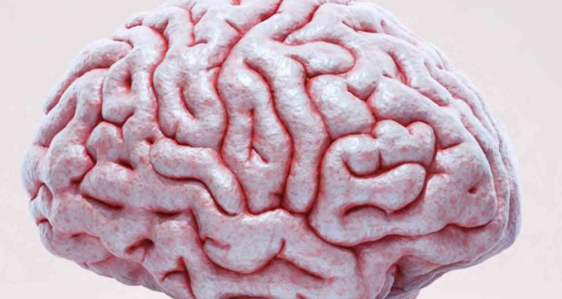
Researchers have developed a way to make living brain tissue temporarily transparent, allowing deep neurons to be seen without disrupting their normal function.
That breakthrough opens the door to watching hidden brain activity more clearly, then returning to the same tissue as it returns to normal.
Clearer views below

In mouse brain slices and living mice, living brain tissue turned visibly clearer within an hour and exposed signals that had been difficult to capture.
By tracking those deeper signals, Takeshi Imai at Kyushu University showed that the cleared tissue still preserved the biology needed for neurons to keep working.
The gain did not last permanently, which was intentional, because the tissue later returned to its usual opacity.
That reversibility makes the result especially striking, and sets up the harder question of how the team achieved it without harming the cells.
Why tissue scatters
Brain tissue looks cloudy because neighboring materials bend light by slightly different amounts, sending rays off course.
Researchers call that light-bending property the refractive index, a measure of how strongly a material redirects light.
Scientists can clear fixed tissues with harsh chemicals, but living cells fail when those solutions pull water or ions out of balance.
That constraint turned the challenge from simple transparency into a harder demand: make tissue clearer without changing how cells behave.
Balancing cells and light
Keeping cells alive required osmotic balance, the water pressure that keeps cells from swelling or shrinking, while nudging tissue toward optical uniformity.
Large molecules helped because fewer of them were needed to bend light, reducing the chemical crowding that can dehydrate cells.
In the final formula, the team chose albumin because that blood protein is highly soluble and already common in circulation.
With albumin in place, the researchers created a solution that cleared tissue, preserved firing, and washed out within hours.
Slices stay active
In brain slices kept alive outside the body, the solution cleared tissue fast enough for repeated imaging sessions.
When researchers added a calcium indicator, a dye that brightens as cells handle calcium, deep neurons kept showing normal firing patterns.
Views in the cortex and hippocampus extended roughly twice as deep with confocal and two-photon microscopy – a laser method for deeper imaging.
That extra reach matters in slices because the uppermost tissue often gets damaged during cutting, while healthier cells sit below.
Testing in live brains
Inside living mice, the team applied the solution through a small skull opening so it could reach tissue beneath the surface.
Because the protein entered cerebrospinal fluid – the liquid that bathes the brain – signals from deep cortical neurons became far brighter.
Lower branches and even tiny protrusions along them came into view about 0.02 inch (0.5 millimeters) below the surface.
Hours later, normal circulation diluted the reagent and the tissue clouded again, making the effect temporary rather than permanent.
Signals still behave
Clearer pictures would mean little if the treatment warped firing, movement, or sensation in the same animals.
Across brain slices and live mouse cortex, neuronal firing and responses to sights and smells stayed largely intact after clearing.
Awake mice also kept normal treadmill movement, food intake, and grip-test performance, suggesting no obvious short-term or longer-term harm.
“This is the first time tissue clearing has been achieved without altering its biology,” said Imai.
Wider biological uses
Beyond brains, the method also cleared cell spheroids and organoids, lab-grown mini tissues that mimic parts of real organs.
Signals that once faded near the surface remained visible much farther in, while growth stayed normal during brief daily treatments.
In intestinal and cortical organoids, cells still responded to stimulation, showing that the clearer view did not erase function.
That makes the reagent useful beyond neuroscience, especially where drug developers need three-dimensional tissues they can watch repeatedly.
Microscopes gain reach
Better transparency did more than brighten pictures because it expanded which microscopes could track fast activity inside living tissue.
With voltage imaging, a method that tracks changes in electrical charge, researchers caught the electrical spikes neurons send along single branches.
Confocal microscopes, which are more common than specialized deep-imaging systems, could now record healthy activity from deeper slice regions.
That broader access could lower costs for labs and speed experiments that once demanded slower or more complex setups.
Ten years later
This result capped a long campaign that began with SeeDB, a chemical method that makes preserved brain tissue transparent, for fixed tissue in 2013.
Three years later, SeeDB2 pushed fixed-sample imaging deeper by matching cleared tissue to high-resolution optics.
“That question came to me about a hundred times, and each time I answered ‘impossible,’” said Imai.
The new live version does not end that story, but it turns a decade of doubt into a practical tool.
Next steps ahead
By matching light behavior outside cells without knocking physiology off course, SeeDB-Live puts chemistry and microscopy into a workable balance.
Researchers still need surgical access to the brain, and other organs remain harder targets, but deeper repeat imaging now looks far more practical.
The study is published in Nature.
—–
Like what you read? Subscribe to our newsletter for engaging articles, exclusive content, and the latest updates.
Check us out on EarthSnap, a free app brought to you by Eric Ralls and Earth.com.
—–