

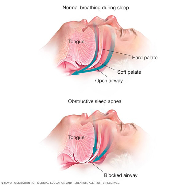
Australian researchers found that CPAP therapy was associated with slower progression of retinal vascular narrowing in severe obstructive sleep apnea. Although RNFL thinning was similar between groups, the findings suggest treating nocturnal hypoxia may have lasting effects on retinal microvasculature and provide insight into broader cardiovascular health. Photo: Mayo Clinic. Click image to enlarge.
Severe obstructive sleep apnea (OSA) has been associated with retinal vascular changes; however, some research has shown that use of continuous positive airway pressure (CPAP) attenuates some of these issues. In one study, an Australian research group demonstrated progressive retinal arterial narrowing in severe untreated OSA, whereas treatment with CPAP appeared to reduce retinal arterial narrowing at a two-year follow-up. The team recently published the results of a seven-year prospective study in the same cohort to reveal whether this protective effect was maintained. The findings appeared in Investigative Ophthalmology & Visual Science.
Of the original 79 participants at the two-year follow-up, 39 individuals were eligible for inclusion in the seven-year study. This included nine with severe OSA plus CPAP, eight with severe OSA without CPAP, three with mild/moderate OSA plus CPAP, 14 with mild/moderate OSA without CPAP and five controls. The mean age was 68 years, and the cohort was 66% male. Assessments were made using OCT and fundus imaging to analyze retinal nerve fiber layer (RNFL) thickness, central retinal artery equivalent, central retinal vein equivalent and arteriovenous ratio.
According to the results, central retinal artery equivalent and central retinal vein equivalent appeared greater in patients with severe OSA plus CPAP than untreated OSA. Over seven years, central retinal vein equivalent declined more slowly in those with severe OSA plus CPAP than those with untreated severe OSA. Severe OSA plus CPAP had a notably smaller decline in central retinal artery equivalent compared with the control and untreated groups.
RNFL thickness was not significantly reduced in severe OSA without CPAP compared with those with CPAP in the superotemporal sector. Global RNFL thinning rates were equivalent across groups. There was a trend for those not on CPAP to have higher rates of hypertension and stroke.
The proposed mechanism for retinovascular changes due to recurrent apneas and hypopneas in OSA is thought to be two-pronged, involving intermittent hypoxia and sympathetic nervous system dysfunction, the authors explained in their paper. “It is proposed that acute hypoxia triggers peripheral chemoreceptor activity, which increases sympathetic nervous system activity, causes episodic systemic hypertension and results in long-term structural changes in both the retinal and systemic vasculature.”
The researchers addressed the limitations within their study, including the loss of nearly half of the original cohort (partially due to the pandemic), as well as pooling of the mild/moderate OSA groups, the potential for undetected OSA in the control group and the use of a 90-day compliance snapshot, as opposed to continuous seven-year monitoring. However, they noted that some participants who were currently noncompliant had used CPAP for a few years before this study. “This may have also had some level of effect on our measurements, actually reducing the differences seen between the groups on analysis,” the team pointed out. “Therefore,” they noted, “the effect of CPAP may be stronger than our data can show.”
The authors concluded that these findings support the hypothesis that CPAP therapy improves hypoxia and reduces retinal vascular narrowing in severe OSA, making it a relevant measurable predictor of cardiovascular health. “Similar to our baseline study and two-year follow-up, this study could guide further evidence gathering and provide data for follow-up studies that are looking at oculomic assessment of retinal vessels, serving as an indirect indicator of systemic cardiovascular risk in participants with OSA,” they wrote.
Click here for the journal source.
This article was developed by the editorial staff in conjunction with experts in the field. In the process, AI may have been among the editorial tools used to meet the goals of human editors, who approved all content.