
Cell lines and cell culture
Bone marrow-derived mouse macrophages were immortalized using the J2 retrovirus, according to the protocol established by Roberson and Walker (1988)74. The growth conditions for iBMDMs (kindly provided by Zeinab Abdullah) were Dulbecco´s modified Eagle´s medium (DMEM) (Thermo Fisher Scientific, #10566016) supplemented with 10% (v/v) fetal bovine serum (FBS) (Thermo Fisher Scientific, #10500064) and 100 U/ml penicillin-streptomycin (Thermo Fisher Scientific, #15140122); the growth conditions for the RAW-Blue NF-κB SEAP reporter cell line (Invivogen, #raw-sp) were the same and in addition 100 µg/ml zeocin (InvivoGen, #ant-zn-05), and 100 µg/ml normocin (InvivoGen, #ant-nr-05).
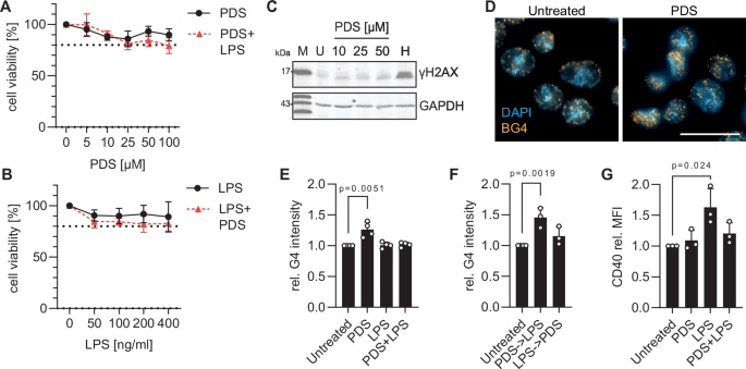
If not indicated otherwise, the cells were treated with 25 µM PDS (Selleckchem, #S7444) and 200 ng/ml LPS (Sigma, #L7770) or H2O2 (30% (w/w) Sigma, #H1009) for 4 h or with 10 µM Nigericin (InvivoGen, #tlrl-nig-5) for 1 h at 37 °C with 5% CO2.
AlamarBlue assay
The metabolic activity was determined using the alamarBlue Cell Viability Assay Reagent (VWR, #GENO786-921). 60,000 cells were seeded in a black bottom clear 96-well plate and 10 µl of 10% alamarBlue solution was added (final volume 100 µl per well) and incubated for 3 h at 37 °C. The emission at 590 nm was measured using a Fluorskan TM FL plate reader (Thermo Fisher Scientific).
Western blot analysis
Cells were treated with PDS, LPS or both for 4 h. Nigericin was added for 1 h to provide the second signal for full inflammasome activation. For detection of secreted proteins, 1 ml of supernatant was precipitated with trichloroacetic acid, washed with acetone, and the pellet resuspended in 30 µl Lämmli buffer. The protein lysates were separated by 10% SDS-PAGEs and blotted on a Protran BA83 Nitrocellulose membrane (GE Healthcare). Blocking was performed with 5% (w/v) BSA (AppliChem, #A6588) in TBS-T and incubated with the primary antibodies overnight at 4 °C with agitation. The membranes were washed three times for 5 min with TBS-T, incubated with appropriate fluorescent-coupled secondary antibodies for 1 h at RT and followed by three washing steps. Reactions were detected using the Molecular Imager ChemiDOC XRS Imaging system (Bio-Rad) with automatically calculated detection times by the ChemiDOC software. ImageJ (Fiji75) was used to determine the protein intensities.
Used antibodies
Phospho p65 (1:1,000 Cell Signaling, #9936); Donkey anti-rabbit 800 CW (1:10,000 LI-COR, #926-32213); Donkey anti-mouse 800 CW (1:10,000 LI-COR, #926-32212); p65 (1:300 Santa Cruz, #sc8008); Phospho-histone H2A.X (1:1,000 Cell Signaling, #9718); GAPDH (1:1,000 Santa Cruz, #sc69778); histone H3 (1:1,000 Abcam, #Ab1791); IL-1β (1:1,000 Proteintech, #26048-1-AP); RelB (1:1,000 Proteintech, #25027-1-AP); Phospho-RelB (1:1,000 Cell Signaling, #5025 T); p105/p50 (1:1,000 Proteintech, #14220-1-AP); Phospho-p50 (1:200 Santa Cruz, #sc-271908); Caspase-1 (1:1,000 Adipogen, #AG-20B-0042-C100); NLRP3 (1:5,000 Proteintech, #30109-1-AP)
Detection of G4 with BG4 antibody
Immunofluorescence staining of G-quadruplex (G4) structures was performed as previously described76. Briefly, immortalized bone marrow-derived macrophages (iBMDMs) were seeded onto glass coverslips in 24-well plates at a density of 7 × 10⁴ cells per well. Twenty-four hours post-seeding, cells were treated with PDS for 4 h. To preserve G4 structures, cells were pre-fixed using a 1:1 mixture of culture medium (DMEM) and methanol/acetic acid (3:1) for 5 min at room temperature. Cells were then incubated in methanol/acetic acid (3:1) for 10 min at room temperature. The fixed cells were permeabilized with 0.1% (v/v) Triton X-100 in PBS for 3 min. Samples were blocked by incubating the coverslips in blocking solution (2% (w/v) nonfat dry milk (Carl Roth, #T145) in PBS, pH 7.4) for 1 h at room temperature. For specific G4 detection, samples were incubated with 2 µg of the FLAG-tagged BG4 antibody per slide in blocking solution for 2 h at room temperature. The primary detection was performed using a rabbit anti-DYKDDDDK epitope antibody (1:800; Cell Signaling Technology, #2368) for 1 h, followed by incubation with a Cyanine 3-conjugated goat anti-rabbit IgG secondary antibody (1:1000; Life Technologies, #A10520) for 1 h at room temperature under gentle rocking. After each incubation step, the cells were washed three times for 10 min with 0.1% (v/v) Tween-20 (Carl Roth, #9127) in PBS. Coverslips were mounted onto glass slides using Fluoroshield mounting medium containing DAPI (Merck, #F6057) for nuclear counterstaining.
BG-flow
BG-Flow was performed as described in DeMagis et al.76. After trypsinization, cells were harvested by centrifugation. Cell pellets were resuspended in 1 ml of a 1:1 mixture of DMEM and methanol/acetic acid (3:1) for 5 min at room temperature (RT). After centrifugation at 300 g for 5 min at RT, cells were fixed in 3:1 (v/v) methanol/acetic acid for 10 min at RT, and then for 15 min at RT. Following two PBS washes, cells were permeabilized with 0.1% (v/v) Triton X-100 in PBS for 5 min at RT and washed twice with PBS. Cells were blocked using 2% (w/v) nonfat dry milk in PBS for 45 min at RT. For target detection, cells were sequentially incubated at RT on a tube rotator (30 rpm) with 5 μg BG4 in blocking buffer for 2 h, followed by a rabbit anti-FLAG antibody (Cell Signaling, #2368; 1:250) for 1 h, and an Alexa Fluor 488 secondary antibody (Invitrogen, #A11008; 1:600) for 1 h. Between antibody incubations, cells were washed twice with 0.1% (v/v) Tween in PBS. After the final incubation, cells were washed once with 0.1% Tween in PBS and once with standard PBS. Where indicated, DNA was co-stained using 10 μg/ml DAPI (alternatively 50 μg/ml PI or 1.2 μg/ml Hoechst33258) and 50 μg/ml RNase A (Ambion, #AM2271) in PBS for 30 min at 37 °C. Finally, cells were resuspended in 1 ml PBS and analyzed on a FACSCanto II Cell Analyzer (BD Biosciences). Data was processed using FlowJo (BD Biosciences), gating populations by forward scatter (FSC) and side scatter (SSC). Samples omitting the BG4 primary antibody served as negative controls.
CD40 and CD80 expression analysis and mitochondrial fitness
The iBMDMs were collected, washed and resuspended in 50 µl antibody mix 1) MTG + MTDR (Fc-block, 1:100, Invitrogen, #14-0161-86; MTG-FITC, 1:4,000, Invitrogen, #M7514; MTDR-APC, 1:4,000, Invitrogen, #M22426; F4/80 BV421, 1:200, #Biolegend 123132; L/D Near-IR, 1:800, Invitrogen, #L34976A); 2) MTG+MitoSox (Fc-block 1:100; MTG-FITC 1:4,000; MitoSox PE, 1:4,000, Invitrogen, #M36008, F4/80, 1:200; L/D, 1:800); 3) (Fc-block, 1:100; CD40 PE, 1:200, eBioScience, #12-0401-83; CD80 AF647, 1:200, Biolegend, #104718; F4/80, 1:200; L/D, 1:1,000) and proceeded for flow cytometry analysis using a FACS Canto I. Appropriate single stains and FMOs were included. FlowJo software was used for analysis.
RNA isolation
The RNA was isolated using TRIzol (Thermo Fisher Scientific, #15596026) according to the manufacturer´s instructions.
RNA sequencing
The 3´RNA sequencing was performed at the NGS core facility Bonn at HiSeq 2500 V4 (10 M raw reads; 1×150 bp). The analysis was performed using the Partek Flow software (Partek Inc.). Sequencing reads were aligned to mm10 using the STAR aligner.
Standard quantitative PCR
For reverse transcription, the QuantiTect Reverse Transcription Kit (Qiagen, #205311) was used according to the manufacturer’s instructions. The quantification of cDNA was performed using the iQ SYBR Green Supermix (Bio-Rad, #170-8880) on a CFX96 Real Time System with a C1000 Touch Thermal Cycler (Bio-Rad) with the primers in Table 1.
Table 1 qPCR primers used for Il1b cDNA quantificationPhagocytosis uptake assay with Salmonella
1×10⁶ iBMDMs were mixed with 50 µl/ml normal mouse serum and incubated for 15 min at 37 °C. 2×10⁸ CFU/ml mCherry-labeled heat-inactivated Salmonella (provided by the Abdullah lab) were incubated for 2 h at 37 °C under rotation. The cells were collected, washed with PBS and stained for L/D (1:800; Near-IR, Invitrogen, #L34976A), CD11b (1:400; FITC, Pharmingen, #553310), F4/80 (1:400; BV421, #Biolegend 123132), Fc Block (1:100, Invitrogen, #14-0161-86) and incubated for 30 min at 4 °C. The samples were washed with PBS and proceeded for flow cytometry analysis using a Fortessa II (BD Biosciences). Appropriate single stains and controls without bacteria were included. FlowJo software was used for analysis.
Phagocytosis killing assay with Listeria
500,000 iBMDMs were seeded (DMEM without antibiotics), if indicated, treated for 20 h with 100 ng/ml recombinant mouse INFγ and infected with 50 µl Listeria (MOI 5). After 30 min the medium was replaced with medium containing gentamycin (100 µg/ml) and after 1 h (t = 0) and 4 h (t = 1) the cells were washed twice with PBS and lysed with 500 µl lysis buffer (0.01% (v/v) Triton X-100 in ddH2O). Serial dilutions (1:10 dilution was diluted 5 times) with the lysate were performed and plated on BHI plates (20 µl were plated). The next morning, the colonies were counted, and the CFU/ml was calculated.
HTRF IL1B
The supernatants of triplicates were collected and the assay performed according to the manufacturer’s instructions (Cisbio, #62MIL1BPEG). In short: 8 µl sample, 8 µl medium, 4 µl antibody mix and 16 µl standard were used, and a SpectraMax M5e plate reader (Molecular Devices) was used (665 and 620 nm).
QUANTI-Blue assay with RAW NF-κB cells (InvivoGen)
The assay was performed based on the manufacturer´s instructions. In brief, 100,000 cells per well were seeded in a 96-well plate. 190 µl QUANTI-BlueTM Solution (InvivoGen, #rep-qbs) were incubated with 10 µl treated RAW-Blue cell supernatant and incubated at 37 °C for 6 h. The SEAP levels were measured using a SpectaMax M5e spectrophotometer at 630 nm.
Procartaplex
The supernatants of the iBMDMs were collected in triplicate. The Procartaplex 9 Plex (Thermo Scientific, #D4445144) was used according to the manufacturer´s instructions. The concentrations were calculated using Thermo Scientific software based on standard curves and measuring the Mean Fluorescence Intensity (MFI).
Inflammasome activation measurement via hASC-mCerulean
Immortalized mouse macrophages with hASC-mCerulean reporter were used for this assay. The well imaging plate (Greiner bio, #6055300) was coated with 0.01% Poly-L-Lysine solution (Sigma-Aldrich, #P8920-100ML) for 15 min at 37 °C and washed three times with PBS. 50,000 (n = 1) and 30,000 (n = 2, 3) cells per well were seeded. The cells were treated with PDS and/or LPS and in the last hour with 10 µM nigericin (Invitrogen, #N1495). In the last 30 min of incubation 50 µM VX-765 solution (Selleckchem, #S2228) was added to all wells. The fixation and nuclear strain were performed in 4% PFA and DRAQ5 (Thermo Fisher Scientific, #62251) (dilution of 1:5000) for 15 min at RT in the dark. The plate was centrifuged at 340 g for min, the supernatant was removed and replaced with 200 µl PBS. For analysis, the automated imaging on the Cell Discoverer 7 (Carl Zeiss Microscopy) was used (imaging settings: 10 images per well, 6 z-stack planes spanning 10 µm with intervals of 2 µm; Objectives n = 1: Plan-Apochromat 20x/0.95 with 1x Tubulens (20x magnification); n = 2,3: Plan-Apochromat 5x/0.35 with 2x Tubulens (10x magnification). The maximum intensity projections from all planes in Zen 2.6 (blue edition, Carl Zeiss Microscopy) software were generated, and the bright field channel was removed. The new file was used for automated quantification in the CellProfiler (3.1.9) Software.
ATAC sequencingSample preparation and sequencing
For ATAC-Seq, 50,000 cells per condition were washed with PBS and lysed in 50 µl of lysis buffer (10 mM Tris-HCl, pH 7.4, 10 mM NaCl, 3 mM MgCl2, 0.1% (v/v) NP-40). Following centrifugation (500 g for 10 min at 4 °C), the nuclear pellet was resuspended in a mix comprising 25 µl of 2x TD buffer, 2.5 µl of Tn5 transposase (Nextera DNA Library Kit, Illumina, #FC-121-1030), and 22.5 µl of nuclease-free H2O. The reaction was incubated for 30 min at 37 °C. DNA was subsequently purified using the MinElute Reaction Cleanup Kit (Qiagen, #28204) according to the manufacturer’s instructions and eluted in 10 µl of elution buffer. Sequencing was performed at the Cologne Center for Genomics on an Illumina platform, using 50 million 150-bp paired-end reads per sample.
Data processing and peak calling
Sequence read preprocessing and alignment were performed using the following pipeline. Raw reads were trimmed using Cutadapt (v1.18)77 and aligned to the mouse reference genome (mm10) using Bowtie2 (v2.4.5)78. Alignments were sorted with SAMtools (v1.14)79 and PCR duplicates were removed using Picard (v2.26.10) (https://github.com/broadinstitute/picard). Peak calling on the sorted and filtered BAM files was performed using MACS2 (v2.2.7.1)80. To ensure high reproducibility across replicates, consensus peaks for each condition were defined using the Irreproducible Discovery Rate (IDR) framework (v2.0.4.2)81. Differentially accessible regions between conditions were identified utilizing the DiffBind package (v3.10) (http://bioconductor.org/packages/release/bioc/html/DiffBind.html).
Fast ChIP
Chromatin immunoprecipitation was performed using the FastChIP protocol established by Bomsztyk and colleagues82. Briefly, iBMDMs were plated at a density of 6–8 × 10⁶ cells in 10 cm dishes. Cells were washed and covered with 5 ml PBS before crosslinking with 1.42% formaldehyde (Carl Roth, #4980) for 15 min at room temperature. The reaction was quenched with 125 mM glycine for 5 min at room temperature, and cells were washed twice with cold PBS, scraped into 1 ml PBS, and pelleted at 2000 g for 5 min at 4 °C. Pellets were resuspended in 1 ml IP buffer (150 mM NaCl, 50 mM Tris-HCl, pH 7.5, 5 mM EDTA, 0.5% NP-40, and 1% Triton X-100, protease inhibitors), and lysed by pipetting. After centrifugation at 12,000 × g for 1 min at 4 °C, the pellet was washed once with 1 ml IP buffer and centrifuged again. For sonication, pellets were resuspended in 1 ml IP buffer per 10 million cells, split into 300 µl aliquots, and sheared using a Bioruptor (Diagenode) for five cycles of 30 s on/30 s off, yielding average fragment sizes of 300–600 bp. Lysates were clarified at 12,000 × g for 10 min at 4 °C, and chromatin aliquots equivalent to 2 million cells were incubated overnight at 4 °C with a 1:100 dilution of a rabbit anti-NF-κB p65 antibody (D14E12, Cell Signaling Technology, #8242S) or an equivalent amount of a rabbit IgG control (Thermo Fisher Scientific, #02-6102. The next day, samples were centrifuged at 12000 g for 10 min at 4 °C, and the supernatant was then incubated with 20 µl of pre-washed protein G-Dynabeads (Invitrogen, #10446293) for 45 min at 4 °C. Beads were washed five times with 1 ml cold IP buffer and resuspended in 100 µl Proteinase K buffer (100 mM Tris-HCl, pH 7.5, 150 mM NaCl, 12.5 mM EDTA, 2% SDS, and 5 mM DTT). Samples were treated with 1 µl of 20 mg/ml Proteinase K (Carl Roth, #3719) for 30 min at 55 °C, followed by boiling at 95 °C for 10-15 min to inactivate the enzyme and decrosslink the chromatin. DNA was precipitated with 300 µl ethanol and 15 µg GlycoBlue (Thermo Fisher Scientific, #AM9515) at −20 °C for at least 1 h, pelleted at 20000 g for 30 min at 4 °C, washed with 70% ethanol, and resuspended in 50 µl water. qPCR was performed with 2 µl of the eluted DNA with the iQ SYBR Green Supermix (BioRad) using the primers in Table 2.
Table 2 qPCR primers used for Fast ChIPAnalysis of NF-κB binding sites and G4s in the Il1b promoter
We analyzed the DNA sequence for NF-κB binding sites using position weight matrices (PWMs) from the HOCOMOCO v11 (https://hocomoco14.autosome.org/) database (RELA, RELB, REL, NFΚB1, NFΚB2) and TFBIND. Both DNA strands were scanned, and for each subsequence, the raw PWM score was normalized to a relative score between 0 and 1 using the theoretical minimum and maximum possible scores for that PWM. Sites with a relative score ≥ 0.80 were retained. To avoid redundancy, predicted sites located within 5 bp of each other were merged by keeping the highest-scoring site in the cluster. G4s were predicted by G4RNA screener83 using the algorithms cGcG, G4hunter and G4NN with a window size of 30 and step size of 5. Running scores are reported for each step.
G4-DNA precipitation (G4DP)
For pulldown of chromatin G4s, a modified variant of the G4DP method established by Feng et al. (2022)51 was employed. Briefly, iBMDMs were seeded at 6 to 8 million cells in 10 cm dishes. The following day, cells were washed, overlaid with 5 ml PBS, and crosslinked using 1% formaldehyde for 10 min at room temperature. Crosslinking was quenched with 125 mM glycine for 5 min at room temperature, followed by two cold PBS washes, scraping into 1 ml cold PBS, and pelleting at 2000 g for 5 min at 4 °C. Cell pellets were lysed in 1 ml cold nuclear isolation buffer (10 mM Tris-HCl pH 7.4, 10 mM NaCl, 3 mM MgCl2, 0.1% NP-40, 1 mM DTT, PMSF) by pipetting and 15 min incubation on ice. After centrifugation at 1000 g for 5 min at 4 °C, nuclear pellets were resuspended in 300 µl sonication buffer (10 mM Tris-HCl, pH 7.4, 5 mM EDTA, 150 mM KCl, 0.5% NP-40, 0.5% SDS, 0.5 mM DTT, PMSF). Chromatin was sheared using a Bioruptor for 10 cycles of 30 s on/30 s off to generate fragments of 100-300 bp, clarified at 12000 g for 10 min at 4 °C, and diluted with 1.2 ml incubation buffer to reduce the SDS concentration below 0.1%. The lysate was treated with 15 µl of 10 mg/ml RNase A for 30 min at 37 °C with shaking. 10% of the lysate was reserved as the input sample. For the pulldown, chromatin aliquots equivalent to ~2 million cells were incubated overnight at 4 °C with 100 µM BioCyTASQ (Merck, #SCT246). Streptavidin Dynabeads M280 (Invitrogen, #10202983) were washed with incubation buffer, added to the sample, and rotated for 4 h at 4 °C. To reduce genomic background, beads were washed twice with 1× rCutSmart buffer (NEB, #B6004S), resuspended in 100 µl of the same buffer with 2 U double-strand-specific dsDNase (Sigma Aldrich, #EN771), and incubated for 10 min at 37 °C with shaking, followed by three washes with cold incubation buffer. DNA was eluted twice using 200 µl elution buffer containing 100 mM NaHCO3 and 1% SDS at 65 °C for 15 min, pooled, supplemented with 16 µl 5 M NaCl, and decrosslinked for 4 h to overnight at 65 °C. DNA was extracted using phenol-chloroform-isoamyl alcohol, precipitated overnight at −20 °C with 15 µg glycoblue and 1 ml ethanol, washed with 70% ethanol until SDS was dissolved, and eluted in 100 µl water. qPCR was performed with 2 µl of the eluted DNA with the iQ SYBR Green Supermix (BioRad) using the primers in Table 3.
Table 3 qPCR primers used for G4DPGeneration of nuclear lysates from iBMDMs
Nuclear extracts were prepared utilizing an optimized rapid fractionation protocol84. Briefly, 5 × 10⁶ cells were harvested, washed with 1 ml of ice-cold PBS, and pelleted. The cell pellet was resuspended in 5 packed cell volumes of hypo-osmotic lysis buffer (0.3 M sucrose, 2% Tween-40, 10 mM HEPES-KOH pH 7.9, 10 mM KCl, 1.5 mM MgCl2, 0.1 mM EDTA, 0.5 mM DTT, protease inhibitors). To release intact nuclei, the suspension was subjected to mechanical disruption via repeated pipetting ( ~ 100 times). The resulting lysate was carefully layered over a 1.5 M sucrose cushion (1.5 M sucrose, 10 mM HEPES-KOH pH 7.9, 10 mM KCl, 1.5 mM MgCl2, 0.1 mM EDTA, 0.5 mM DTT, and protease inhibitors) and centrifuged for 10 min to isolate the nuclear fraction. The nuclear pellet was washed with low salt wash buffer (10 mM HEPES-KOH pH 7.9, 10 mM KCl, 1.5 mM MgCl2, 0.1 mM EDTA, 0.5 mM DTT, and protease inhibitors) and subsequently extracted in 100 µl of high salt extraction buffer (20 mM HEPES-KOH pH 7.9, 420 mM NaCl, 1.5 mM MgCl2, 0.2 mM EDTA, 25% glycerol, 0.5 mM DTT, and protease inhibitors) for 20 min on ice with intermittent gentle vortexing. The extract was cleared via centrifugation, and the supernatant containing high-purity nuclear proteins was collected, aliquoted, and stored at −80 °C.
Electrophoretic mobility shift assay (EMSA)Oligonucleotide hybridization and G-quadruplex folding
Fluorescently labeled oligonucleotides were designed to contain a central G-quadruplex (G4)-forming motif flanked by duplex-forming genomic sequences. To facilitate structured G4 folding within the flanking double-stranded context and to mimic the transient single-stranded state during transcriptional activation, a specialized denaturation-renaturation sequence was applied. Sense and antisense strands were diluted to a working concentration of 0.6 µM in binding buffer (10 mM Tris–HCl pH 7.4, 1 mM EDTA, 150 mM KCl, and 5% glycerol). The mixtures were denatured, flash-cooled on ice, and subsequently allowed to fold and anneal at 37 °C in the presence or absence of 25 µM PDS. Untreated control duplexes and single-stranded references were prepared under identical conditions. As a positive control, a double-stranded oligo containing an NF-κB consensus sequence (Cy5-GTTGAGGGGACTTTCCCAGGCA) that does not fold into a G4. This sequence was also used as an unlabeled competitor oligo to validate the NF-κB-dependency of shifted bands.
The sequences of the used oligos are shown in Table 4.
Table 4 Oligonucleotides used for EMSAIn vitro binding and electrophoretic separation
EMSA binding reactions were performed in a total volume of 18 µl. Each reaction mixture contained 1× binding buffer, 1 µg of poly(dI:dC) (Thermo Fisher Scientific, #20148E), and 1–2 µl of purified nuclear lysate. For competition controls or super-shift experiments, a 100-fold excess of unlabeled consensus oligonucleotide was included during a pre-incubation phase of 5 min at 20 °C. Then, 2 µl of the pre-folded fluorescent target oligonucleotide was added, followed by an additional incubation for 30 min at 20 °C. Protein-DNA complexes were resolved on a 5.5% native TBE polyacrylamide gel. The gel was pre-run on ice for at least 30 min at 120 V in 0.25x TBE running buffer. Samples were separated at 180 V for approximately 40 min until the bromophenol blue migration front reached the lower third of the gel. Fluorescent signals were visualized by direct gel imaging on a BioRad Chemidoc system. Quantification was performed on composite images (Cy3 + Cy5) using Fiji.
Reporting summary
Further information on research design is available in the Nature Portfolio Reporting Summary linked to this article.