
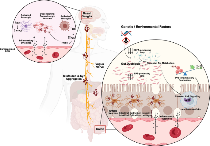
Parkinson’s disease (PD) is the second most common progressive neurodegenerative disease worldwide, inflicting health and economic burden on society1,2. This debilitating disease affects almost 11 million people around the world, with the number of cases predicted to double in the near future3. PD has been classically known as a motor disorder featuring the degeneration of dopaminergic neurons in the substantia nigra pars compacta (SNpc), and characterized by symptoms like gait issues, bradykinesia, resting tremors, and muscle stiffness4. However, this view has been reshaped in recent decades due to the consistent reporting of early non-motor symptoms in patients with PD, such as sleep disorders, loss of sense of smell, and gastrointestinal issues such as constipation5,6,7. This new insight highlights the fact that PD is a multidimensional systemic disorder and implicates the emerging role of gut-brain axis in PD pathology. Gut-brain axis is a bidirectional communication between the central and enteric nervous systems (Fig. 1), modulated directly via vagus nerve, or indirectly through the bioactive metabolites produced by the commensal gut bacteria8.
Fig. 1: Hypothetical alterations of gut-brain axis in Parkinson’s disease.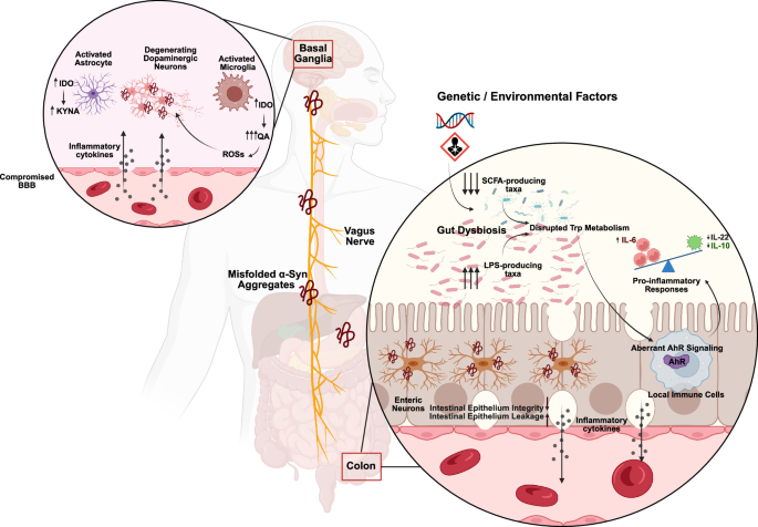
Schematic representation of the bidirectional communication between the gastrointestinal tract and the brain in Parkinson’s disease (PD), with a focus on the role of tryptophan (Trp) metabolism and microbiota-derived metabolites in the pathology of the disease. The vagus nerve serves as a bridge linking the gut to the brain. In the colon, gut dysbiosis is characterized by a reduced abundance of beneficial short-chain fatty acid (SCFA)-producing and AhR ligand-producing commensals (e.g., Lactobacillus, Clostridium, Bacteroides), alongside an overgrowth of lipopolysaccharide (LPS)-producing pathobionts. This microbial imbalance disrupts Trp metabolism, leading to diminished production of anti-inflammatory indole derivatives and an imbalanced kynurenine pathway, where neurotoxic quinolinic acid overrides the protective effects provided by kynurenic acid. The resulting dysregulation of AhR signaling shifts the immune tone toward heightened pro-inflammatory and reduced anti-inflammatory activity. Collectively, these cellular disturbances contribute to compromised gut barrier integrity (leaky gut), local inflammation, and α-synuclein misfolding within enteric dopaminergic neurons.
A compelling line of evidence supports the gut-to-brain transmission of the disease due to the spread of gut-originated misfolded alpha-synuclein (α-syn) aggregates to the other tissues9,10,11. α-syn is a naturally occurring pre-synaptic protein involved in regulation of synaptic neurotransmission12. Under disease conditions (Fig. 1), α-syn can form oligomeric fibrils, promoting the misfolding and aggregation of this protein13,14. Aggregated α-syn fibrils then spread and seed the other post-synaptic neurons in a prion-like manner15. The idea of gut-first PD was initially introduced by Braak et al., suggesting that PD pathology starts in periphery and gradually transmits to the central nervous system via the vagus nerve16.
Central to this gut-brain axis is the metabolism of an essential amino acid, tryptophan (Trp)17. Upon digestion, Trp is metabolized through three major metabolic pathways: serotonin, kynurenine, and indole pathways (Fig. 2). These metabolic routes, particularly the kynurenine and indole pathways, give rise to a wide range of important bioactive metabolites that contribute to the immune signaling and integrating endogenous and environmental cues18,19,20.
Fig. 2: Tryptophan metabolic pathways and aryl hydrocarbon receptor (AhR) ligands in the gut-brain axis.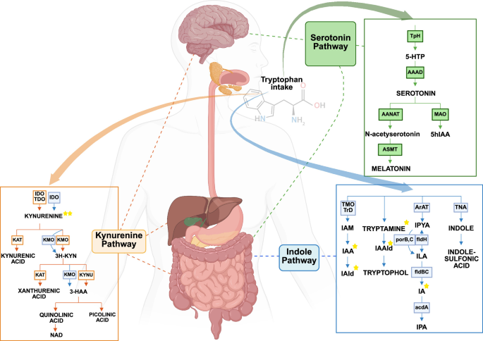
Schematic representation of the three major branches of tryptophan (Trp) metabolism in the human body, kynurenine, serotonin, and indole pathways, and their spatial relevance across gut, liver, and brain compartments. Trp metabolism is initiated by both host and microbial enzymes, generating a diverse array of metabolites with distinct biological functions. The kynurenine pathway (orange box), primarily initiated by indoleamine 2,3-dioxygenase (IDO1/IDO2) and tryptophan 2,3-dioxygenase (TDO), produces several neuroactive and immunomodulatory metabolites, including kynurenine (KYN), kynurenic acid (KYNA), and quinolinic acid (QUIN). Enzymes written in blue represent bacterial enzymes, highlighting the partial microbial contribution to this pathway. The indole pathway (bottom right, blue box) is exclusively microbial, involving the transformation of Trp by gut bacteria into AhR-active indole derivatives such as indole-3-acetic acid (IAA), indole-3-aldehyde (IAld), and tryptamine. The serotonin pathway (top right, green box) converts Trp into 5-hydroxytryptophan (5-HTP) and serotonin (5-HT) via host enzymes such as tryptophan hydroxylase 1 (TPH1), largely occurring in the gut. Metabolites marked with a star (★) indicate known AhR ligands.
Progressive degeneration of serotonergic terminals has been established in PD, associated with both motor and non-motor symptoms of the disease21. Interestingly, some studies have shown that the serotonergic neurons of the raphe nuclei are often affected by PD-associated pathological changes early in the disease, occasionally predating dopaminergic degeneration22,23,24. This early onset of serotonergic dysfunction may explain the prodromal non-motor symptoms of PD, such as depression, apathy, and impaired motivated decision-making22,23,25. As dopaminergic neurons progressively degenerate, spared serotonergic terminals take up exogenous L-DOPA and convert it to dopamine, which can further contribute to the development of motor dysfunctions. These serotonergic neurons release the synthesized dopamine in a non-physiological manner that initiates L-DOPA-induced dyskinesia26,27,28. Although some serotonergic modulators, such as citalopram or 5-HT1A agonists, may help alleviate these involuntary dyskinetic movements, they can also pose risks, such as serotonin syndrome27,29,30.
Although serotonin is well known as a neurotransmitter in the brain, ~95% of serotonin is synthesized by the enterochromaffin cells in the gut31, likely providing a crucial connection between Trp metabolism, gastrointestinal physiology, and CNS function32. In addition to its classic role in gut motility33, serotonin is also known to have additional functions in the gut, some of which may be regulated by aryl hydrocarbon receptor (AhR)34. AhR is a ligand-activated transcription factor that serves as a molecular sensor and mediates many crucial physiological functions, from glial activation to immune signaling and barrier integrity35. In fact, serotonin can interact with AhR-regulated xenobiotic enzymes, such as CYP1A1, thereby indirectly modulating AhR activity through influencing the clearance of its other high-affinity ligands36. Future studies are required to decipher the interactions of serotonin and AhR signaling in the context of PD and its associated gut dysbiosis.
In this review, we report the mechanistic contributions of the gut-brain axis in PD and provide evidence linking gut dysbiosis, disrupted Trp metabolism, with a particular focus on the kynurenine and indole pathways, and altered AhR signaling to the neuroinflammation observed in PD. This review is an attempt to elucidate the multifaceted pathology of PD, providing a framework for understanding potential biomarkers of early PD.
Overview of AhR structure and signaling pathwaysMolecular structure of AhR
AhR is a member of the basic helix-loop-helix-periodic circadian protein (PER)-AhR nuclear translocator (ARNT)-single-minded protein (SIM) (bHLH-PAS) superfamily of transcription factors37. Human AhR is 848 amino acids long and its molecular structure comprises of four predominant domains: an N-terminal bHLH domain modulating DNA binding and protein-protein interactions, including dimerization; PAS-A and PAS-B domains, the former facilitating dimerization and the latter modulating ligand binding processes; and lastly, a C-terminal transactivation domain (TAD) involved in transactivation machinery recruitment38 (Fig. 3). Inactive AhR is a part of a cytoplasmic chaperone complex encompassing heat shock protein 90 (HSP90), co-chaperones like X-associated protein 2 (XAP2, also known as AIP), p23, and the tyrosine kinase c-Src39. This complex contributes to AhR stability in the cytoplasm and prevents its degradation. A conformational change resulting from ligand binding enables AhR to detach from the chaperone complex, interact with different transcriptional or non-transcriptional partners, that vary depending on the ligand and cell type40. Although evolutionary well conserved, AhR ligand specificity and downstream outcomes depend on the organism, cell, and tissue type41.
Fig. 3: Structural domains of the aryl hydrocarbon receptor (AhR).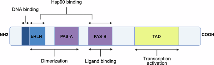
Schematic representation of the functional architecture of the AhR protein, a ligand-activated transcription factor belonging to the bHLH-PAS family. The protein contains four principal domains: bHLH (basic helix-loop-helix): Involved in DNA binding and protein-protein interactions with other transcriptional partners. PAS-A and PAS-B (PER-ARNT-SIM domains): Critical for dimerization and ligand binding, respectively. TAD (Transactivation domain): Located at the C-terminal end, responsible for recruiting the transcriptional machinery necessary for target gene expression.
AhR is known for its role in detoxification and metabolism of xenobiotic ligands such as dioxins (e.g., TCDD) and polycyclic aromatic hydrocarbons42. However, currently, it is characterized as a pleiotropic regulatory receptor, mainly as a sensor and integrator of a wide range of environmental cues, endogenous, and microbial signals, including the metabolites from Trp metabolism such as kynurenines and indoles43.
In humans, different indole compounds, including those synthesized through microbial Trp metabolism in the gut, can function as AhR agonists or antagonists, depending on the context. For instance, some recent evidence suggests that slight methylation or methoxylation modifications in indoles determine the pharmacological effects of these compounds; therefore, 4-methylindole acts as a potent agonist, whereas 3-methylindole functions as an antagonist44. Notably, the presence of strong ligands like dioxin might alter the functional behavior of certain agonists toward antagonistic effects.
Mechanistically, the structural plasticity of AhR, which stems from diverse ligand-binding modes, provides AhR with high functional versatility. This functional multimodality establishes the foundation for its contribution to central and peripheral biological processes in health and disease (Fig. 4).
Fig. 4: Pleiotropic roles of the aryl hydrocarbon receptor (AhR).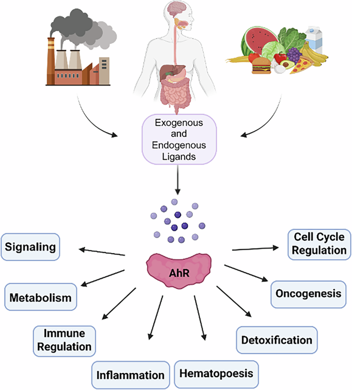
Schematic illustration of the central and multifaceted nature of the AhR as a signaling hub responsive to a wide range of exogenous (e.g., environmental toxins) and endogenous (e.g., dietary and microbial metabolites) ligands.
Canonical AhR signaling
Canonical AhR signaling is a well-established signaling pathway, wherein ligand activated AhR translocates to the nucleus and heterodimerizes with AhR nuclear translocator (ARNT)45. This nuclear translocation is followed by binding of the AhR-ARNT heterodimer to a set of specific DNA motifs, called xenobiotic-/dioxin-responsive elements (XRE/DRE), in the promoter region of different target genes. Upon this DNA binding, required co-activators and transcriptional machinery are recruited to this site, initiating the transcription of the target gene46 (Fig. 5).
Fig. 5: Canonical, non-canonical, and non-genomic signaling pathways of AhR.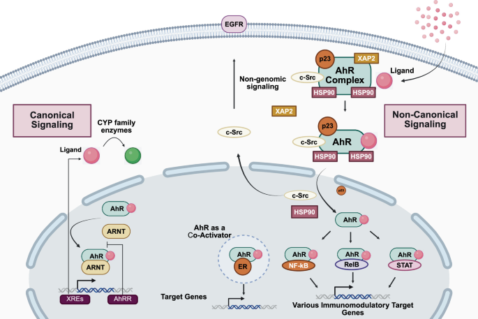
This figure illustrates the diverse signaling routes of the Aryl Hydrocarbon Receptor (AhR). In the canonical pathway, ligand-bound AhR translocates into the nucleus, dimerizes with ARNT, and activates gene transcription at xenobiotic response elements (XREs). Non-canonical signaling involves interactions with other transcription factors (e.g., NF-κB, STATs) independent of XRE binding. Non-genomic pathways include rapid cytoplasmic signaling via kinases such as c-Src and functions such as ubiquitination and proteostasis regulation, highlighting the multifaceted roles of AhR in immunity, metabolism, and inflammation.
Amongst the best-studied target genes in canonical AhR signaling are the members of the cytochrome P450 (CYP) family, including CYP1A1, CYP1A2, and CYP1B1 genes. As a result of the expression of these genes, xenobiotic enzymes are produced, which catalyze the metabolism of many endogenous and exogenous compounds47,48,49. Another set of genes expressed via the canonical signaling, are genes like AhR repressor (AHRR) and TCDD-inducible poly ADP-ribose polymerase (TIPARP), both of which provide a negative feedback regulatory response to suppress AhR activity50,51.
Non-canonical AhR signaling pathwayGenomic functions and transcriptional crosstalk
In addition to these well-characterized canonical pathways, AhR contributes to a broad range of physiological functions independent from ARNT, either through transcription regulation or cytoplasmic events. These pathways are referred to as non-canonical AhR signaling pathways, providing AhR with additional versatility and emphasizing its importance in health and disease (Fig. 5). It has been shown that ligand-activated AhR can collaborate with the NF-κB signaling pathway, particularly through the RelB subunit, to synergistically induce the production of IL-22 in toll-like receptor (TLR)-activated macrophages in the bone marrow52. This is an example of non-canonical contribution of AhR in immune responses and cytokine regulation. Another example of XREs-independent regulation of gene expression by AhR is the regulatory complex that it forms with the Signal Transduction and Activator of Transcription 1 (STAT1), which attenuates the inflammation triggered by bacterial lipopolysaccharide (LPS)53. This complex can suppress the transcriptional activity of NF-κB at the promoter region of IL-6, reducing the inflammatory response. Furthermore, AhR engages in distinct crosstalk with multiple nuclear receptors, with distinct outcomes based on the ligand concentration for each receptor, time, and also cellular contexts. For instance, there is a bidirectional relationship between AhR and the estrogen receptor (ER)54. This relationship often displays as a reciprocal inhibition, where agonists of each receptor can decrease the transcription of the other receptor’s target genes. However, under some exceptional circumstances, AhR ligands can temporarily increase the transcription activity of the ER. Additionally, AhR and glucocorticoid receptor (GR) demonstrate a bidirectional crosstalk in some cells, like human retinal pigment epithelial cells55. In this case, GR activation reduces gene expression mediated by AhR; in contrast, AhR activation enhances GR-mediated gene expression. Moreover, AhR can also interact with the retinoic acid receptors (RARs), either synergistically or in an inhibitory manner. For example, one study has shown that co-treatment with agonists of both receptors can synergistically induce the expression of matrix metalloproteinase-1 in keratinocytes, but conversely inhibiting AhR-responsive genes like CYP1A156.
Collectively, these non-canonical signaling pathways broaden the functional capacity of AhR, granting its ability to integrate several environmental, metabolic, and immune signals in a context-dependent fashion; pathways that progressively appear to be relevant to the pathology of multiple neurodegenerative disorders such as PD.
Non-genomic functions
Besides the previously discussed transcriptional functions such gene expression mediation, AhR appears to engage in pivotal non-genomic and cytoplasmic activities that regulate diverse essential signaling pathways. This additional cytoplasmic contribution of AhR in sub-cellular processes solidifies its association with the pathology of different diseases.
One well-defined non-canonical role of AhR is its ability to function as an E3 ubiquitin ligase. This ligase activity regulates protein stability in a ligand-dependent manner57. In this mode, AhR contributes directly to ubiquitination machinery and promotes proteasomal degradation of selected target proteins, including steroid hormone receptors. This mechanism allows AhR to modulate estrogen and androgen receptor signaling through post-translational control rather than transcriptional interference, establishing a rapid and reversible regulatory framework that bridges environmental cues to endocrine signaling.
A second major class of non-genomic AhR activity involves cytosolic kinase signaling, particularly through interactions with Src-family kinases. In this context, AhR activation promotes Src-dependent phosphorylation cascades that influence cell adhesion, migration, and proliferation with no transcriptional activation required. For instance, AhR-Src interaction has been shown to regulate focal adhesion dynamics via downstream activation of focal adhesion kinase (FAK), thereby linking environmental or endogenous ligands to cytoskeletal organization and cell motility through a non-genomic pathway58. In line with this kinase regulatory function, another study shows that TCDD activated AhR activates cytoplasmic Src kinase. Activated Src kinase, in turn, mediates the release of ligands that activate the epidermal growth factor receptor (EGFR). Activated EGFR, subsequently, activates the ERK1/2 pathway, that increases cell proliferation59.
Non-genomic AhR signaling is also evident in immune cells, where it mediates inflammatory responses through post-translational regulatory mechanisms rather than transcriptional activity. For example, AhR activation in endotoxin-activated macrophages modulates different cytoplasmic events required for innate immune signaling without requiring a direct gene expression induction60. In parallel, AhR can influence cytokine-driven transcriptional responses indirectly by altering the dynamics of transcription factor binding. For example, research has shown that AhR regulates the monocytic responses to IL-4, not through a direct interaction with the DNA, but via prolonging STAT6 binding duration to the target promoters. This increased binding time results in substantial shifts in functional program and metabolic fate of the monocytes in response to IL-4, proving the non-genomic contribution of AhR to the immune responses61.
Finally, functional versatility of AhR further extends to angiogenic response regulation through both non-genomic and genomic mechanisms62. This functional diversity and the complex signaling framework of AhR, beyond mediating gene expression, underline the role of AhR as an extremely flexible regulatory molecule, which explains its mounting recognition in the scientific literature as a significant factor associated with multiple inflammatory and neurodegenerative diseases like PD.
The Indole-Kynurenine-AhR axis in health and PDImmune and barrier homeostasis
As discussed previously, AhR is a ligand-activated transcription factor that converts environmental, dietary, microbial, and endogenous chemical signals into tissue-specific transcriptional programs. Upon activation, AhR regulates xenobiotic-response genes, while interactions with other transcription factors enables AhR to mediate a broad range of physiological functions. For instance, its crosstalk with NF-κB allows it to modify inflammatory transcription63,64. One of the key physiological roles of such signaling is at barrier surfaces, where AhR ligand availability reflects dietary and microbial metabolism. Alterations in dietary AhR ligands reshape the composition and function of the gut microbiota. Altered ligand availability also modulates AhR-repressor expression in intestinal immune cells65,66. AhR therefore functions not merely as a sensor, but as a bidirectional interface through which the host and gut microbiota interact and reciprocally influence one another.
In the intestinal epithelium, AhR controls the structural integrity and permeability of the barrier. For example, epithelial AhR tightly regulates Wnt-β-catenin signaling in intestinal stem cells thereby supporting proper barrier regeneration following inflammatory injury67. Additionally, in some models of barrier injury, AhR has been shown to preserve tight junction stability and restrain epithelial permeability. Several microbial Trp metabolites, including indole-3-ethanol, indole-3-pyruvate, and indole-3-aldehyde, maintain the apical junctional complexes in an AhR-dependent manner68. Moreover, indole-3-carboxaldehyde activates the AhR-AMPK signaling, which supports autophagy, mitochondrial function, and junctional integrity69. Furthermore, AhR sustains mucosal barrier through mediating the normal Muc2 expression, independently of IL-2270. IAA also promotes protective mucin sulfation, partially through AhR71.
Barrier integrity and immune homeostasis are inseparable; AhR activation through dietary ligands maintain intestinal intraepithelial lymphocytes72, while ILCs produce IL-22, restrict segmented filamentous bacteria, and constrain expression of the pathogenic Th17 cell, all via activating AhR73. Similarly, microbial I3A induces AhR-dependent of IL-22 production74, whereas IAcryA provides epithelial protection and inflammation suppression75. In addition, AhR promotes the synthesis of IL-10 by regulatory B cells76, and limits macrophage pyroptosis through polyamine biosynthesis77. AhR also enhances xenobiotic efflux at the BBB78 and induces anti-inflammatory or reparative responses in astrocytes and microglial cells79,80,81. However, AhR ligand availability and transcriptional partners, including NF-κB, determine these effects63. For instance, KYN can alleviate colitis82, whereas exercise-associated AhR/IDO changes can alter NK cell function83. Consequently, disrupted AhR signaling may weaken epithelial and immune defenses, creating a self-reinforcing cycle of barrier leakage84, dysbiosis, altered ligand production, and inflammation.
This homeostatic framework may also be relevant to PD (Table 1), where dysbiosis can alter Trp metabolism and, consequently, the repertoire of host- and microbially derived AhR-active metabolites.
Table 1 Summary of PD-associated AhR mechanisms
Altered or aberrant ligand signaling may weaken the mucosal barrier and tight junctions, resulting in increased intestinal permeability (leaky gut) that allows microbial products, or other toxic compounds to enter the circulation and amplify cytokine production. Consequently, these circulating inflammatory mediators can affect the BBB and resident glia103,104,105. Importantly, these events are not necessarily linear, as ligand identity, concentration, exposure duration, and each cell type can each elicit distinct AhR responses ranging from protective and compensatory to pro-inflammatory.
There is evidence from PD-models supporting a microbiota-AhR-inflammation connection. In a study utilizing rotenone-induced rat PD models, dietary Trp improved motor function, preserved nigral tyrosine hydroxylase, reduced serum levels of IL-1β, IL-6, and TNF-α, and inhibited NF-κB activation. Intriguingly, ampicillin treatment weakened these effects, and the AhR antagonist CH223191 abolished them84. In an MPTP mouse PD model, AS-IV remodeled microbiota, restored indole-3-carboxaldehyde, and suppressed microglial NLRP3/caspase-1 activation, α-syn accumulation, dopaminergic loss, and motor impairment; fecal transfer reproduced protection, although the involvement of AhR signaling remains to be elucidated106. Anthocyanin treatment likewise changed indole and KYN metabolites while reducing neuroinflammation and preserving dopaminergic markers; however, its proposed link to AhR signaling remains associative, requiring further AhR-focused investigations107. Importantly, in a study of patients with PD the plasma levels of IPA were significantly elevated108, a metabolite that has been previously shown to induce mitochondrial dysfunction109 and prolonged AhR activation with potential oncogenic effects110.
Several other findings indirectly support a potential link between AhR signaling and PD pathology. Administration of Enterococcus hirae QT4713, a dopamine-producing strain, restored goblet cells, colonic expression of ZO-1 and claudin-1, while lowering intestinal and systemic inflammation, and protecting nigral neurons in an MPTP mouse model of PD111. In another study, dopamine stimulated AhR signaling in colon-derived and glioblastoma cells86; however, the microbial dopamine-AhR connection remains inferential in other cases, including PD, due to cell type-specific nature of these results. Furthermore, in a rotenone mouse PD model, IPA treatment offered intestinal and BBB protection and reduced enteric glial activation via PXR and IL-13Rα1-JAK1-STAT6 signaling, rather than directly through AhR signaling112. Moreover, the AhR-active microbial metabolite 1,4-DHNA improved MPTP-associated motor deficits more effectively than an inactive isomer, suggesting potential AhR involvement113.
Within the brain, AhR signaling may determine whether glial activation promotes neuroprotection or neurotoxicity. For instance, in LPS-stimulated microglia, ligand-free AhR activity promoted the expression of inflammatory markers iNOS, COX-2, TNF-α, and neurotoxicity, whereas addition of AhR ligands shifted signaling toward anti-inflammatory effects89. In an MPTP mouse model of PD, MPTP dynamically increased activated AhR in nigral and striatal microglia and striatal astrocytes. This glial activation was proposed to contribute to neuroinflammation, while in surviving dopaminergic neurons, sustained AhR activation appeared to be neuroprotective88. AhR also transactivates the parkin promoter, resulting in increased parkin expression and reduced α-syn levels, which may connect AhR to mitochondrial quality control and proteostasis85. However, persistent toxicant-driven AhR activation may instead promote oxidative stress and inflammatory brain aging114.
Collectively, the evidence supports dysregulated, rather than uniformly excessive or deficient, AhR signaling in PD. Disturbed microbial metabolism can erode barrier-supportive signaling, amplify peripheral inflammation, and alter glial responses, while neuronal AhR may preserve parkin-dependent defenses. Disease outcome therefore appears to be context-dependent, depending on which ligand activates AhR, in which tissue and cell type, and for how long.
Kynurenine and indole metabolism in health and PD: host and microbial contributions
Kynurenine pathway (KP) takes place mainly in the liver (~95%); however, it is also modulated by the central or peripheral immune cells of the host and to a lesser extent by the gut microbiota115. Kynurenine (KYN), kynurenic acid (KYNA), and quinolinic acid (QA) are examples of the metabolites with neuromodulatory functions, with KYNA acting as a neuroprotective116 and QA as a neurotoxic metabolite117. While KP occurs in both host and bacterial cells, indole pathway is broadly known as a gut microbiota-exclusive metabolic pathway118,119, through which important indole derivatives are produced including indole lactic acid (ILA), indole acetic acid (IAA), indole propionic acid (IPA), and indole-3-carbaldehyde (I3A). Maintaining the balance of these pathways is crucial in health. Growing evidence suggests that disruptions in Trp metabolism may contribute to development, progression, and exacerbation of various neurodegenerative diseases, including PD108,120,121.
Many of the metabolites generated via kynurenine and indole pathways can act as ligands for AhR122,123,124. Given its functional variability, AhR is implicated in the pathogenesis of many diseases125, from cancer126 to neurodegenerative disorders like PD105. In PD, gut dysbiosis disrupts Trp metabolism, which leads to altered levels of kynurenine and indole metabolites, resulting in aberrant AhR signaling. Dysregulated AhR signaling, in turn, results in chronic central and systemic inflammation127, one of the hallmarks of PD pathology128. For instance, imbalanced KP metabolites have been frequently observed in many PD cases, characterized by reduced levels of KYNA and increased QA levels, an excitotoxicity regulator and a neurotoxic metabolite, respectively129.
Host-driven kynurenine metabolic pathway
Trp metabolism is the predominant immunometabolic node, where host and gut microbiome enzymatic activities interface to integrate the endogenous and exogenous metabolic cues and provide a flexible framework conducive to maintaining the homeostatic environment within the host organism. These reciprocal interactions produce many bioactive metabolites involved in signaling through receptors, including AhR. The kynurenine pathway (KP) is the key Trp metabolizing route in mammals, through which nearly 95% of dietary Trp is catabolized to generate multiple neuroactive metabolites115,130. Thus, a strictly regulated and balanced KP is essential for the physiology and health of an organism131,132. Dysfunctional KP has been observed as an underlying factor in many human diseases, particularly neuroimmune disorders such as PD131,133 (Table 2). For instance, a recent study has reported significantly increased levels of KYN and the neurotoxic 3-hydroxykynurenine (3-HK) in the cerebrospinal fluid of the patients with PD. Notably, a number of KP metabolites, as shown in Fig. 6, can interact with AhR98,134,135. These interactions potentially modulate AhR activity and its associated immune signaling.
Fig. 6: Hypothetical implications of peripheral and central kynurenine pathway metabolism in immune-neural crosstalk.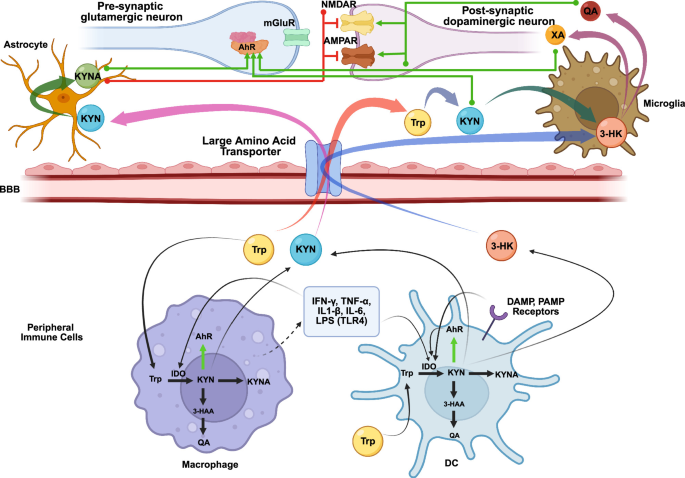
This schematic illustrates the compartmentalized metabolism of tryptophan (Trp) through the kynurenine pathway (KP) across peripheral immune cells and central nervous system (CNS). In peripheral tissues, hepatic TDO and extrahepatic IDO initiate Trp degradation into kynurenine (KYN), which can cross the BBB. Within the CNS, KYN is further metabolized by astrocytic KATs to neuroprotective kynurenic acid (KYNA) or by microglial KMO to neurotoxic quinolinic acid (QA). These metabolites exhibit distinct effects on NMDA receptor signaling, KYNA acting as an antagonist and QA as an agonist, thus critically influencing the neuroprotective-neurotoxic balance. In the context of PD, the overall effect appears to reflect toxic QA outweighing the neuroprotective effects of KYNA. KYN, generated from various host and bacterial cells, also functions as an endogenous AhR ligand, modulating transcriptional responses in both immune and barrier tissues.
Table 2 Summary of PD-associated Trp metabolism
Additionally, balance between KYNA and QA production is of great importance since they are an NMDA receptor antagonist and agonist, respectively115,131,132,136 (Fig. 6). Therefore, KYNA can provide neuroprotective support against QA-driven excitotoxicity137. Upregulated KP towards the neurotoxic branch, elevates 3-HK and QA, promoting oxidative stress, which leads to neuroinflammation130,131,132,133. Recent research provides compelling evidence of the new therapeutic potentials through manipulating KP138. Inhibiting kynurenine 3-monooxygenase (KMO), a key enzyme that produces neurotoxic 3-HK and QA, has been shown to successfully shift KP towards the production of the neuroprotective KYNA137. Moreover, some studies, administering KMO inhibitors that can cross the BBB, observed restored balance in KYNA and QA production, which may stop or slow neurodegeneration130.
Gut microbiota, dysbiosis, and AhR ligand availability
Besides the host-driven KP, the indole pathway is another key route of Trp metabolism (Table 2). This pathway is carried out exclusively by the gut microbiome (Table 3) and is responsible for metabolizing almost 10% of dietary Trp124. Gut commensal bacteria, such as Escherichia coli and Clostridium sporogenes, convert Trp into various indole derivatives, including indole, indole-3-propionic acid (IPA), indole-3-lactic acid (ILA), indole-3-acetic acid (IAA), and indole-3-carboxaldehyde (I3C)98,124,135. These indole derivatives can interact with and activate AhR; therefore, they are essential for host-microbiota interactions and host physiology. As discussed earlier, several indole metabolites are vital for mucus secretion, antimicrobial-protein production, and tight-junction-protein expression, thereby maintaining gut-barrier integrity. Additionally, this indole-AhR axis is pivotal for IL-22-dependent immune regulation by Tregs and ILC3s98.
Table 3 Gut microbiome and its PD implications
Building upon these findings, the intestinal microbiota performs fundamental roles in immune regulation and barrier homeostasis by metabolizing dietary Trp through the indole pathway and producing essential AhR ligands74. One frequently investigated genus of gut bacteria is Lactobacillus74,97. This genus plays a key role in generating metabolites such as indole-3-carboxaldehyde (IAld) through the indole-3-pyruvic acid (IPyA) pathway. Many microbially produced indole derivatives are necessary for inducing IL-22 secretion, thereby directly contributing to host resistance against mucosal pathogens and the maintenance of epithelial integrity74. Interestingly, specific species such as Lactobacillus reuteri have been shown to facilitate the reprogramming of CD4 + T cells into regulatory intraepithelial lymphocytes by utilizing Trp-derived metabolites and activating AhR97. This broad metabolic activity forms a regulatory interactome in which microbial Trp metabolites participate in host immunomodulation and secure an effective mucosal response.
An imbalanced gut microbiome, known as gut dysbiosis, significantly compromises the availability of AhR ligands and contributes to the pathology of multiple inflammatory and metabolic disorders99,164,165. For example, genetic defects in the caspase recruitment domain family member 9 (CARD9) gene cause gut dysbiosis, thereby hindering the conversion of Trp into indole metabolites such as IAA164. Reduced AhR-ligand availability is also involved in metabolic syndrome, in which decreased AhR activity correlates with impaired glucose metabolism and a high body mass index in both mice and humans165. These alterations in gut-microbiota composition often divert Trp metabolism away from the advantageous indole pathway and toward the host-driven KP99. This metabolic shift can further disrupt intestinal-barrier function and intensify mucosal inflammation.
The bidirectional communication along the gut-brain axis propagates the adverse effects of altered AhR-ligand availability beyond the gut and can influence CNS health92,105. Different studies have shown that several indole metabolites, such as IPA and IAld, can cross the BBB115,166. This brain permeability underlines their immunomodulatory role, enabling these metabolites to regulate glial activity and provide neuroprotection against oxidative stress and amyloid-related toxicity166,167. For instance, in chronic neuroautoimmune conditions, microbially derived Trp metabolites can reach the CNS and activate microglial or astrocytic AhR. This activation results in the downregulation of pro-inflammatory gene expression and suppression of neuroinflammatory responses92,105. In some neuroimmune disorders, such as MS, the levels of these microbial AhR agonists are significantly reduced, and their availability has been inversely correlated with disease duration168. Likewise, as discussed earlier, gut dysbiosis has been reported to alter the levels of aromatic amino-acid metabolites, including indoles99,169,170. Interestingly, administration of natural AhR agonists or specific probiotic strains can restore normal AhR signaling, offering a potential therapeutic avenue for mitigating neuroinflammation, re-establishing proper immune function, and supporting CNS health170,171.
On the other hand, some microbial indole metabolites, such as indoxyl sulfate (IS) and skatole, can be toxic and are regarded as uremic or toxic metabolites172,173. Intriguingly, some beneficial indoles known for their anti-inflammatory effects can become toxic at high concentrations. For example, elevated concentrations of IAA are involved in chronic kidney disease and contribute to cardiovascular and liver dysfunction174. Elevated IAA levels have also been identified as a key neuroinflammatory factor underlying the pathogenesis of neuropsychiatric systemic lupus erythematosus. Excess IAA can cross the BBB and activate microglial AhR/STAT3 signaling, resulting in neural deterioration and cognitive deficits87.
Moreover, some of these metabolites can activate AhR in different tissues, leading to pathological outcomes. A notable example occurs in chronic kidney disease, in which impaired renal clearance leads to systemic IS accumulation, promoting progressive inflammatory responses, oxidative stress, and necrotic or apoptotic cell death175. IS is also known as an endotheliotoxin that exerts cardiovascular damage associated with renal failure172,176. Additionally, IS can interfere with glial function, resulting in the oxidative stress and neuroinflammation observed in chronic kidney disease91. However, these findings conflict with another study investigating the effects of IS on dopaminergic neuronal death and motor function in PD177. The toxic effects of indole metabolites, particularly IS, have not been fully explored in the context of PD, highlighting the need for deeper and more elaborate research. Furthermore, studies have shown that skatole can activate AhR and trigger pro-inflammatory outcomes in the gut. These effects contribute to the disruptions of intestinal homeostasis observed in inflammatory bowel disease and colorectal carcinoma95,96.
Collectively, this interplay between host and microbial Trp metabolism, converging on AhR signaling pathways, forms a dynamic triad that governs immune regulation, mucosal homeostasis, and neuroimmune communication. In PD, the breakdown of this regulatory system is evidenced by changes in serum levels of KP metabolites and altered fecal or plasma levels of microbial indole metabolites108,129,145,178,179,180. These imbalances may disrupt AhR-mediated signaling cascades both in the gut and brain, contributing to chronic inflammation and dopaminergic neurodegeneration. Understanding the distinct but interacting contributions of host- and microbiota-derived Trp catabolites, potentially through AhR signaling, provides a framework for examining how alterations in gut microbial communities further perturb this signaling axis and impact disease progression.
AhR signaling in other neurodegenerative disorders and neuropathologic conditions
Emerging evidence suggests from disorders other than PD suggests that AhR signaling may be a potential underpinning of the pathology of several other neurological conditions in addition to PD. However, similar to PD, AhR signaling cannot be classified as uniformly protective or pathogenic. Instead, its consequences vary with ligand identity, cellular compartment, disease stage, and duration of activation, offering useful parallels for understanding dysregulated AhR signaling in PD.
In Alzheimer’s disease (AD), several studies support a protective role for AhR activation by microbiota-derived indoles. In APP/PS1 mice, indole, IAA, and IPA activated AhR while suppressing NF-κB signaling, NLRP3 inflammasome formation, and the release of TNF-α, IL-6, IL-1β, and IL-18204. These changes were accompanied by reduced amyloid-β (Aβ) accumulation and tau hyperphosphorylation, improved synaptic plasticity, and better cognitive performance. Similarly, microbiome-derived ILA reduced soluble Aβ levels and ameliorated cognitive impairment in 5xFAD mice through AhR-dependent effects involving microglia and astrocytes205. Supporting translational relevance, the same study identified greater abundance of Lactobacillus reuteri in healthy individuals than in individuals with early amyloid accumulation or mild cognitive impairment and detected stronger AhR-associated transcriptional signatures in non-AD postmortem brains. These findings connect microbial Trp metabolism to glial regulation and aggregate clearance, processes also implicated in PD.
Conversely, host KP activation may engage AhR differently in AD. Aβ-treated neurons and APP/PS1 mice displayed altered KP enzymes and increased activity of the IDO1-KYN-AhR axis, whereas IDO1 inhibition attenuated inflammatory responses and partially restored of neurotrophic signaling206. Although the AhR causality is not directly isolated, it may suggest that excessive upstream IDO1 activity and the resulting ligand environment may shift AhR signaling toward neuroinflammation. Thus, even within AD, microbial indoles and host-derived KYN may produce different AhR-dependent outcomes.
Evidence from Huntington’s disease (HD) further supports a potentially pathogenic role for sustained endogenous AhR activity. Genetic deletion of AhR in R6/1 transgenic mice prevented clasping, improved motor coordination and ambulatory behavior, normalized anxiety-related behavior, and reduced astrogliosis207. These findings could particularly be relevant to PD since both disorders involve motor impairment, vulnerable striatal circuitry, pathogenic protein accumulation, and reactive glia. Interestingly, in an ALS-related study, AhR agonists including dioxins, polychlorinated biphenyls, and FICZ increased soluble and insoluble TDP-43 in human induced pluripotent stem cell-derived neurons, and mouse brain by enhancing TDP-43 expression and limiting protein clearance208. Although this was not a complete ALS disease model, it directly links environmentally activated AhR to impaired proteostasis, similar to the accumulation of α-syn in PD.
Stroke studies demonstrate similarly divergent effects. Experimental ischemia increased KYN-dependent AhR activity, suppressed CRE-dependent neuroprotective genes including BDNF and NPAS4, and promoted neuronal injury, while inhibition of TDO or AhR was protective209. AhR activation in microglia also increased the NADPH oxidase component p47phox, oxidative stress, TNF-α expression, lipid peroxidation, and vasogenic edema after middle cerebral artery occlusion210. These mechanisms resemble PD-associated microglial activation, oxidative injury, and loss of trophic support. However, stroke also disrupted the balance between AhR ligands by decreasing microbiota-derived indoles while increasing host-derived KYNs211. Restoring indole-based ligands reduced microglial inflammation, infarct volume, and neurologic deficits in aged mice, indicating that ligand balance, rather than total AhR activation alone, determines outcome.
Depression provides additional evidence linking AhR to systemic immunometabolism. In patients with major depressive disorder (MDD), plasma KYN inversely correlated with symptom severity, while AhR SNP variants were associated with KYN concentrations. Further functional experiments showed that AhR regulates KP enzymes in astrocyte- and hepatocyte-derived cells thereby may contribute to the pathology of MDD212. In an LPS-induced mouse model, FICZ restored hippocampal AhR/CYP1A1 signaling, inhibited NLRP3 inflammasome, reduced IL-1β and IL-6, and alleviated depressive-like behavior213.
Collectively, these studies position AhR as a ligand-, cell-, and disease-dependent integrator of microbial and host metabolism, glial inflammation, oxidative stress, and protein homeostasis, supporting a similarly nuanced role in PD.
Therapeutic potential of the KYN-indole-AhR axis in PD
As discussed so far, mounting evidence supports the key role of gut microbiota and AhR-Trp axis in health and disease, largely due to the fact that bacterial Trp metabolism generates several AhR ligands. Understanding this axis presents potential therapeutic opportunities in PD (Table 4).
Table 4 Summary of potential therapeutic implications of KYN-Indole-AhR axis in PD
Fecal Microbiota Transplantation (FMT) is an emerging PD therapeutic strategy aimed at restoring the healthy gut biome in PD subjects via fecal transfer from healthy individuals. FMT in PD mouse models, where a healthy human microbiome was transferred to mice, has been shown to decrease dopaminergic neuronal loss and partially improve motor functions102. However, these rodent findings are not fully replicable in human FMT studies. For example, a recent 3-year clinical trial (2020–2023), coupled with a 12-month follow-up study on 45 PD patients, demonstrated that FMT, despite overall methodological safety, did not provide clinically meaningful improvements214. Two other very recent pilot studies also showed similar results, where there were no significant objective and sustained improvements between FMT and placebo-controlled groups215,216. Thus, while FMT offers some promise as an adjunctive strategy, further research is required.
Several studies suggest that targeting the AhR-Trp axis through probiotic administration may help restore gut-brain axis function, offering a compelling strategy to mitigate neurodegeneration. For example, specific probiotic strains such as Bacillus subtilis PXN21 have demonstrated the ability to promote clearance of α-synuclein aggregates217. Another study highlights the effects of nutritional indoles, such as indole-3-carbinol (I3C) and 3,3′-diindolylmethane (DIM), in inducing IL-22 production and enhancing tight-junction protein expression, thereby supporting mucosal integrity and immune regulation218. Both ligands have been shown to shift the Th17/Treg balance toward an increased Treg population, which may reduce systemic inflammation, a well-known underlying factor of dopaminergic neurodegeneration. Additional research notes that the microbial metabolite indole-3-aldehyde (3-IAId) activates ILCs to maintain gut epithelial integrity219. Administration of these derivatives may help restore barrier function in the context of PD-associated “leaky gut” and prevent translocation of pro-inflammatory mediators into systemic circulation, thereby addressing PD at its proposed enteric origins.
Conversely, modulating Trp intake through dietary restrictions can potentially represent a complex therapeutic strategy for PD. Despite some promising outcomes provided by dietary protein restriction, aimed at reducing the production of uremic toxins in diseases like chronic kidney disease, this approach needs to be carefully balanced to prevent the loss of neuroprotective metabolites, particularly those derived from Trp metabolism. Since Trp is a large neutral amino acid, it can competitively inhibit transport of levodopa across the BBB220,221. As a result, protein-restricted or protein-redistributing diets might improve levodopa bioavailability and motor functions. Moreover, dietary restriction methods have shown compelling therapeutic effects in some autoimmune and neuroinflammatory conditions, including MS222,223,224,225.
Along with diet-focused strategies, repurposing currently available medications can also represent a feasible and cost-effective approach for clinical intervention in PD management. Several approved drugs have been identified as potential candidates, including febuxostat and rabeprazole, which act as AhR agonists capable of restoring gut barrier integrity and significantly reducing Toll-like receptor 4 expression226. Collectively, these findings support the possibility of leveraging existing pharmaceuticals to suppress the sustained systemic inflammation underlying PD pathology. Additional studies highlight the therapeutic potential of anakinra, an AhR activator acting through modulation of the kynurenine pathway227. This mechanism promotes autophagy, restores mitochondrial redox balance, and enhances cellular proteostasis, providing multifaceted protection against oxidative stress and protein misfolding that contributes to neuroinflammation and dopaminergic neurodegeneration.
This therapeutic perspective offers the potential to slow the progression of PD by acting on disease-relevant immunometabolic pathways, complementing conventional dopaminergic therapies that primarily address symptomatic relief rather than underlying pathology. However, it is noteworthy that the success of AhR-targeted therapies, defined by maximum efficacy and minimal off-target effects, requires advanced drug delivery systems, which could be challenging. For instance, the highly context-dependent function of AhR necessitates technologies such as enteric microparticles or nanoparticle systems to achieve site-specific delivery of the administered drugs227,236. Additionally, high inter-individual variability and heterogeneity of the human gut microbiome remain the primary obstacle in developing effective drugs targeting the AhR-Trp axis217,219. Therefore, establishing standardized treatment protocols will require further clinical trials designed around personalized metabolic frameworks.