
Patients’ characteristics
Among the 25 patients, 20 received ECT primarily for symptom palliation and local disease control, whereas in five patients ECT was used with the aim of improving local tumor conditions and facilitating subsequent surgical resection, as determined by multidisciplinary tumor board evaluation. The study group consisted of 14 males (56%) and 11 females (44%), with a mean age of 49.56 ± 13.01 years (Median = 51.0, range: 19–69). The patients’ characteristics, tumor histotype, tumor size, previous treatments, ECT session numbers, and tumor RECIST were shown in Table 1. Regarding the grade of tumors: nine had grade I (36%), while five had grade II (20%), and eleven had grade III (44%). Twenty-three patients presented with tumors ≥ 5 cm in largest diameter (92%), while the remaining 2 (8.0%) had medium lesions (between 3 and 5 cm). Median largest tumor diameter was 87 mm (mean = 103, range 35–320 mm). The tumor sites included the trunk (52%), and upper/lower limbs (48%). Among the participants, 17 (68%) had experienced tumor recurrence, while 2 (8%) had metastatic disease. The metastatic sites included the right scapula and inguinal lymph nodes. 20 patients underwent surgical procedures at least once, with a maximum of five surgeries and a median of two. Radiation therapy was administered for 13 patients (52%), and 11 patients (44%) underwent multiple ChT courses. In addition, two patients received immunotherapy. Two patients were treatment-naïve at the time of multidisciplinary evaluation. In both cases, limb-sparing resection with adequate oncologic margins was considered infeasible or associated with unacceptable functional morbidity because of the extent and anatomical involvement of the tumors, and amputation was recommended by the multidisciplinary tumor board. Both patients declined amputation. Following further multidisciplinary discussion of available treatment options, ECT was offered as an individualized, non-standard locoregional treatment aimed at local disease control, limb preservation, and subsequent reassessment for surgery. All patients with fibromatosis were discussed in the multidisciplinary tumor board before treatment. ECT was considered only in selected patients with recurrent, locally advanced, or symptomatic disease after multidisciplinary evaluation, when previous treatments had failed to provide durable local disease control or when further conventional treatment was considered unsuitable or not feasible because of anticipated functional morbidity, patient-specific considerations, or treatment accessibility. A total of 44 ECT courses were administered (median two per patient, range 1–4). Eleven (44.0%) patients received a single ECT, ten (40.0%) received two ECTs, three (12.0%) received three ECTs, and one (4.0%) patient received four ECTs. The median treatment duration was 25 min (range 10–40), and hospital stay was 1 day (range 1–4).
Table 1 The patients’ & tumors characteristics.Pain & quality of life
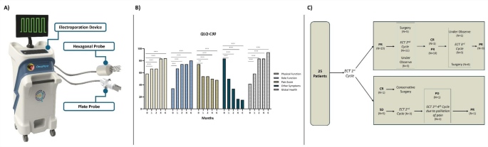
Overall symptom palliation was achieved in 16 of 18 symptomatic patients (88.9%). Among the 14 patients with severe baseline pain, 12 (85.7%) experienced a reduction in VAS score after ECT, with individual reductions ranging from 66% to 100%. Bleeding and tumor-associated discharge resolved completely in all eight affected patients.
Quality-of-life analysis demonstrated significant improvement across all evaluated EORTC QLQ-C30 domains following ECT (Fig. 1). The mean Global Health score increased from 41.66 ± 7.00 at baseline to 93.73 ± 8.13 at the 6-month follow-up, representing a mean improvement of 52.07 points (95% CI, 48.87–55.27; P < 0.001). Physical Function improved from 58.33 ± 10.70 to 83.98 ± 15.21, with a mean improvement of 25.65 points (95% CI, 21.45–29.84; P < 0.001). Similarly, Role Function increased from 34.00 ± 13.41 to 80.00 ± 13.91, corresponding to a mean improvement of 46.00 points (95% CI, 42.56–49.44; P < 0.001). Conversely, symptom burden significantly decreased, with the Pain score decreasing from 74.00 ± 8.55 to 48.00 ± 11.80 (mean reduction, 26.00 points; 95% CI, 22.08–29.92; P < 0.001) and the Other Symptoms score decreasing from 83.33 ± 8.60 to 15.00 ± 12.67 (mean reduction, 68.33 points; 95% CI, 65.24–71.42; P < 0.001).
Local tumor control
Final local response was assessed two months after the last ECT session in 25 target lesions. Treatment workflow and clinical outcomes are summarized in Fig. 1. Initial radiologic assessment demonstrated one complete response (CR) and 19 partial responses (PRs). Five patients (20.0%) demonstrated stable disease. Among the patients with an initial PR, subsequent management included surgical resection (n = 5), a second ECT cycle (n = 11), or active surveillance for more than 6 months (n = 3). Following additional ECT sessions, one patient achieved CR, resulting in a total of two complete responders. The histological subtypes of the complete responders were fibromatosis (n = 1) and undifferentiated pleomorphic sarcoma (n = 1). One patient who achieved CR subsequently developed local recurrence 5 months after treatment, declined further intervention, and died 7 months later because of progressive pulmonary metastases.
Among the remaining patients with PR, ten maintained partial response after the second ECT cycle. During further follow-up, six patients underwent surgical resection, one remained under observation, and three received a third ECT cycle, resulting in sustained PR in all three cases. In CR/PR cases, tumors showed significant shrinkage, necrotic transformation, and reduced enhancement (Figs. 2 and 3). The median percentage of tumor necrosis on electroporated tumors, assessed on pathological specimens, was 50% (range 20–100).
Fig. 2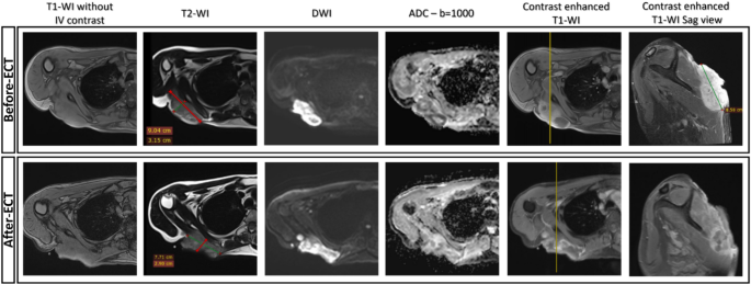
A 47-year-old male with a Dermatofibrosarcoma located posterior to the right scapula. Prior to ECT, the tumor measured 90 × 85 × 31 mm, exhibiting two diffusion-restricted ring-shaped areas surrounding cystic/necrotic regions. Follow-up imaging 60 days post-ECT shows marked tumor size reduction, significantly increased ADC values, and prominent necrotic zones within the lesion. Notably, there is also reduced contrast enhancement.
Fig. 3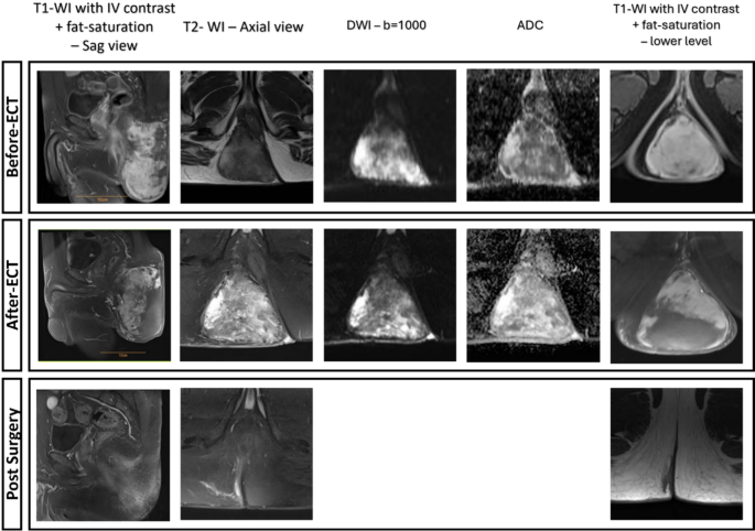
In the first row, the initial pelvic MRI images demonstrate a mass in the right buttock of a 46-year-old man. The hypervascular mass is in close contact with the deep posterolateral right external anal sphincter, with no discernible fat plane between the mass and the anus. Given its morphology, size, restricted diffusion on DWI, and low ADC values, the mass was deemed hypercellular and biopsied, with sarcoma as the primary differential diagnosis. Pathology revealed no necrosis and a mitotic rate of 1–2 per 10 HPF. In conjunction with immunohistochemistry, these findings confirmed a low-grade solitary fibrous tumor. The patient underwent two sessions of ECT, delivered to a depth of 10 cm. The second row displays follow-up images obtained two months after the last ECT session. The mass persists in close contact with the external anal sphincter, still lacking a clear fat plane. A subtle irregularity in the anterior portion, where the mass abuts the sphincter muscle, raises the possibility of local invasion without mucosal involvement. Notably, there is a loss of enhancement in the posterior inferior portion of the mass, along with reduced diffuse enhancement and elevated ADC levels, particularly in the lower sections. Additionally, a necrotic area measuring 101 × 52 mm is evident on contrast-enhanced and diffusion-weighted images. The third row presents post-surgical resection images, showing no residual bulk mass, with only immature fibrotic changes remaining.
Six patients were initially considered candidates for amputation before electrochemotherapy. ECT enabled initial limb preservation in all six patients. Three patients subsequently demonstrated sustained local tumor control without requiring further intervention, while three underwent surgical resection after improved operability. Two of the surgically treated patients later developed postoperative infectious complications and ultimately required amputation. Consequently, long-term limb preservation was achieved in four of the six patients who were initially considered candidates for amputation.
Among the five patients with initial stable disease, one achieved a partial response following additional ECT sessions, three remained progression-free with stable disease for more than 6 months, and one subsequently developed disease progression. Approximately one year after treatment, the patient with PD underwent surgical resection and subsequently required amputation because of postoperative infectious complications.
Finally, an objective response was reported in 21/25 (84.0%, 95% CI: 63.9%-95.5%) patients. As follows, three examples of patients who underwent ECT were shown in Fig. 4.
Fig. 4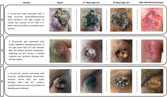
Three examples of patients with STS who underwent ECT.
Local progression free survival & overall survival
After a median follow-up of 13.7 months (mean = 15.26 ± 8.59), 1-year LPFS and OS rates were 56.0% and 76%, respectively. During follow-up, 11 local recurrences had occurred (44%), and five patients developed new lesions outside the ECT field (20.0%), after a median of 8.5 months (95%CI = 4.38–12.61). In patients experiencing disease progression, the median time to local failure was 12.5 months (95%CI = 5.28–19.71).
At the time of analysis, six patients (24%) had died due to systemic disease progression. The mean overall survival was 29.9 months (95% CI: 22.7–37.1 months). The median overall survival was not reached because fewer than 50% of patients experienced the event during the follow-up period.
Toxicity
ECT was associated primarily with local tissue-related adverse effects. The most frequent local adverse event was skin reaction, observed in 24/25 patients (96.0%), including 14 grade 3 events. Soft-tissue necrosis occurred in 12/25 patients (48.0%), including 10 grade 3 events, while soft-tissue infection occurred in 6/25 patients (24.0%). Skin ulceration was observed in 1/25 patients (4.0%), and outpatient surgical debridement was required in three patients (12.0%), with a mean treatment duration of 1 month. No grade 4–5 or serious systemic ECT-related adverse events occurred. Toxicity data are presented in Table 2.
Table 2 Systemic & local toxicity after electrochemotherapy.