

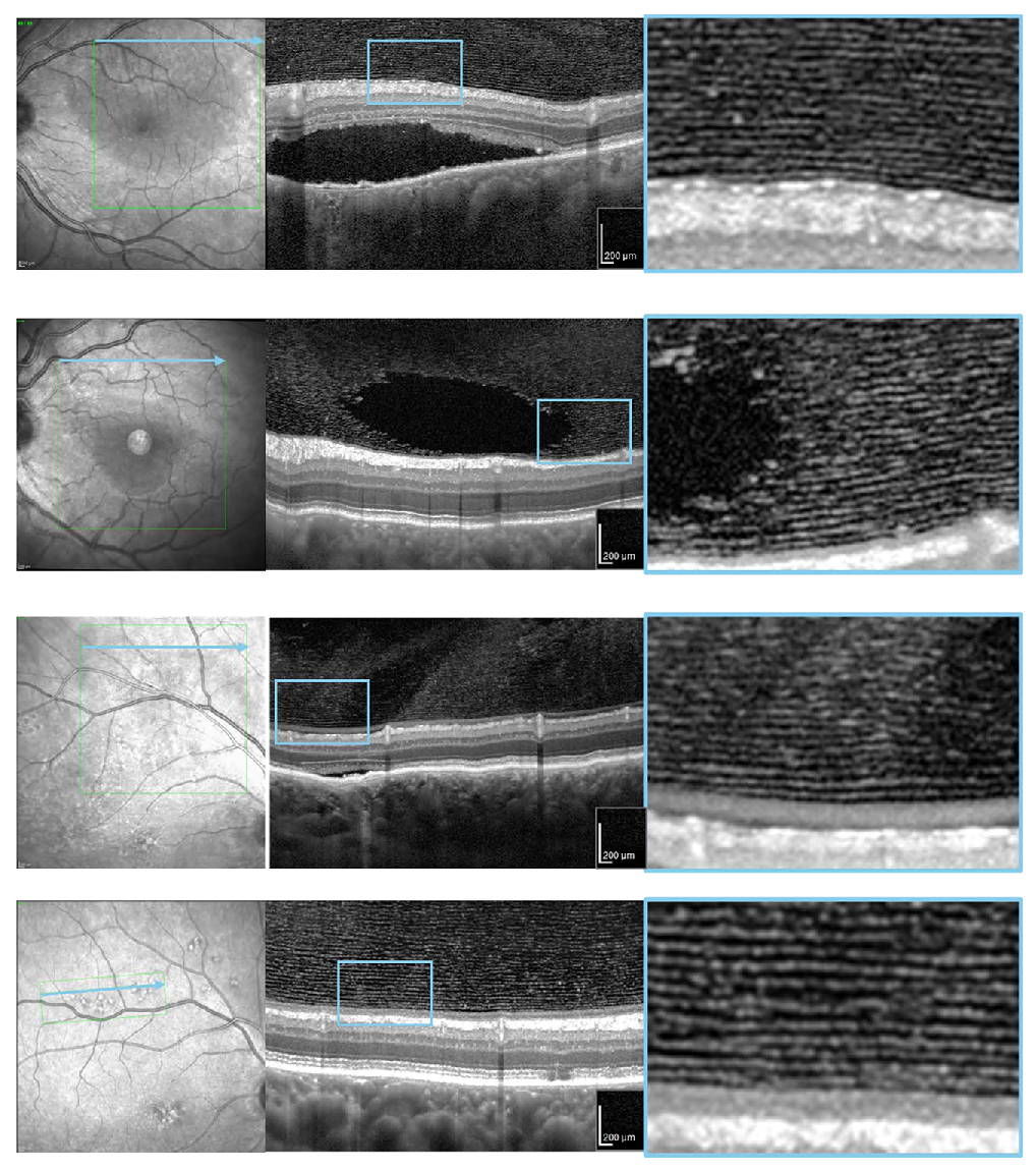
Distinct layered patterns were preferentially observed in eyes with chronic CSCR, suggesting that disease-associated alterations in vitreous matrix organization may increase the optical visibility of collagen architecture. These high-res OCT images from the study show representative cases of four CSC patients. The right column contains magnified images of the area indicated by the square in the central figure. Photo: Miura Y, et al. Translational Vis Sci Tech. 2026;15(9):14. Click image to enlarge.
Changes in collagen organization have been suggested to be associated with aging, oxidative stress, posterior vitreous detachment and retinal disease. However, despite its potential relevance as a sensor of intraocular or systemic molecular alterations, the vitreous has traditionally been viewed as only sparsely structured on clinical OCT, as most of its fibrillar components remain below the resolution of conventional imaging.
Because chronic central serous chorioretinopathy (CSCR) frequently affects relatively younger individuals without advanced posterior vitreous detachment, it may provide a particularly suitable condition for in vivo evaluation of posterior vitreous microarchitecture. Researchers in Germany used high-res OCT, a new Spectralisimaging platform from Heidelberg, to examine posterior vitreous microstructure in patients with chronic CSCR compared with healthy individuals to characterize the visibility and qualitative features of vitreous lamellar structures and to evaluate whether such structures may reflect disease-related alterations in vitreous matrix organization.
The team determined that high-res OCT enabled in vivo visualization of posterior vitreous lamellar microstructures that are not readily discernible with conventional clinical OCT. The preferential detection of these structures in eyes with chronic CSCR suggests disease-associated alterations in vitreous matrix organization and highlights a previously underexplored dimension of vitreous imaging.
“The axial resolution of 3µm enables visualization of subtle lamellar structures within the posterior vitreous that are not detectable with conventional systems,” the study authors wrote in their paper, which was published in Translational Vision Science & Technology. “Conventional SD-OCT has been limited in its ability to image the vitreous, which arises from the low scattering properties of the vitreous combined with the insufficient axial resolution of standard clinical systems.”
High-res OCT imaging (axial resolution approximately 3µm) was performed in 20 patients with chronic CSC and 20 age-matched healthy subjects. Posterior vitreous structures were qualitatively assessed for the presence of discrete lamellar patterns and compared with conventional spectral-domain OCT acquired at the same visits.
Distinct hyperreflective lamellar structures within the posterior vitreous were clearly identified in nine of 20 CSCR eyes, whereas no comparable lamellar organization was observed in healthy eyes. In CSCR eyes, lamellae appeared as parallel, layered bands located immediately anterior to the internal limiting membrane and were reproducible across scan orientations. These structures were not readily discernible on conventional OCT. Healthy eyes showed relatively homogeneous vitreous reflectivity, although faint linear structures were occasionally observed.
“Together, these findings suggest that molecular remodeling of the ocular microenvironment may extend to the vitreous compartment and potentially influence the vitreous matrix,” the researchers noted. “Future studies combining high-res OCT with aqueous humor or vitreous biochemical analyses may help clarify whether the increased visibility of lamellar structures reflects disease-related biochemical or biophysical remodeling of the vitreous matrix.”
Click here for the journal source.
This article was developed by the editorial staff in conjunction with experts in the field. In the process, AI may have been among the editorial tools used to meet the goals of human editors, who approved all content.