
Ethical approval and animal use
All animal procedures were conducted in accordance with the German Animal Welfare Act (Tierschutzgesetz) and with permission from the responsible authority, the Government of Upper Bavaria (ROB-55.2-2532.Vet_02-24-131). Bovine ovaries were obtained from a slaughterhouse (Münchner Schlachthof Betriebs GmbH, Munich, Germany). In vitro produced embryos were obtained from an EU-approved bovine embryo collection and production center at the Chair for Molecular Animal Breeding and Biotechnology of the LMU Munich (Moorversuchsgut Badersfeld, Oberschleißheim, Germany; approval number DE ETR 006 EWG).
Animal experiments utilized cattle (Bos taurus). Primary nuclear donor cells (BFF-091019) were established from an early-gestation female Limousin fetus (B. taurus), and all generated embryos, day-18 conceptuses (n = 4 IFNT-null, n = 3 wild-type), day-50 fetuses (n = 2 IFNT-null), day-75 fetuses (n = 2 IFNT-null), and live-born calves (n = 2 IFNT-null) were female. Sex was not evaluated as a comparative biological variable in the study design or statistical analyses, and sex-disaggregated analysis within or between experimental groups was not applicable. A total of 22 cyclic recipient heifers, primarily of the German Simmental breed (B. taurus) and crossbreds (German Simmental × Holstein-Friesian), were utilized for estrus synchronization and embryo transfers.
Generation of IFNT-KO cells
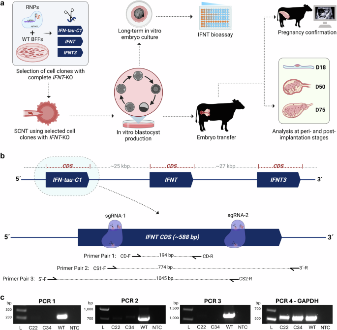
Primary BFF cells, internally designated as BFF-091019, were established from an early-gestation female Limousine fetus (B. taurus) obtained from the slaughterhouse following standard protocol. Cells were maintained under standard culture conditions at 37 °C with controlled humidity and 5% CO₂ in fibroblast growth medium supplemented with 15% fetal calf serum (FCS), non-essential amino acids, and HEPES buffer. The single-guide RNA (sgRNA) design and genomic locus annotation were based on the latest ARS-UCD2.0 bovine genome assembly (NCBI RefSeq assembly GCF_002263795.3; GenBank assembly GCA_002263795.4) submitted by Rosen et al.52,53. The targeted region included all six alleles of the three functional IFNT genes: IFN-tau-c1 (Gene ID: 528153), IFNT2 (Gene ID: 317698), and IFNT3 (Gene ID: 100313956). Multiple sgRNAs targeting all annotated functional IFNT loci were designed by using established in silico tools to ensure high on-target efficiency and minimal predicted off-target effects. Several sgRNA pairs were tested, and the pair demonstrating the highest editing efficiency across all IFNT copies (sgRNA1: catcagtagagagagcacga; sgRNA2: agtgcagagctgctccagga) was selected to generate the final IFNT-knockout (KO) donor cells for SCNT.
For genome editing, pre-assembled Cas9 ribonucleoprotein (RNP) complexes were prepared by mixing TrueCut™ Cas9 Protein v2 (Thermo Fisher Scientific, Waltham, MA, USA) with the selected sgRNAs, and subsequently delivered into BFFs by electroporation with the Amaxa Nucleofector I device. Edited cell populations were allowed to recover for 48–72 h before single-cell sorting and clonal expansion in 96-well plates. Clones were propagated, and genomic DNA (gDNA) was extracted for molecular characterization. Three independent PCR assays targeting distinct regions of the multigene IFNT locus were used to identify candidate knockout clones. Primer Pair 1 amplified a ~200-bp fragment of gDNA within the IFNT coding sequence (CDS) between the two sgRNA cut sites (CD-Forward: tgaggaccacatgctaggtg; CD-Reverse: ggaagaggttgaagcactgc). Primer Pair 2 spanned the gDNA from a forward primer overlapping the sgRNA1 cut site to a reverse primer in the 3′-UTR, yielding a ~774-bp product (CS1-Forward: gtgctctctctactgatggcc; 3′-Reverse: tcagggcaggcatcacttaa). Primer Pair 3 amplified a ~1045-bp gDNA fragment from the 5′-UTR to a reverse primer overlapping the sgRNA2 cut site (5′-Forward: gtgtattccccctccctgag; CS2-Reverse: gtgcagagctgctccagga). GAPDH served as a housekeeping control (F: cgggtgcaattaacttgctcctt; R: cttctgggtggcagtgatgg). Clones lacking amplification across all three IFNT-specific assays, indicative of complete deletion of all IFNT copies, while retaining GAPDH product, were designated as complete knockouts. Starting with the single parental BFF-091019 population, multiple single-cell clones were generated and screened for complete IFNT-KO. Following comprehensive molecular validation, two independent IFNT-KO single-cell clones (SCC-22 and SCC-34) were selected for all subsequent in vitro functional assays and in vivo embryo transfer experiments on the basis of their confirmed complete deletion of all functional IFNT alleles and superior growth characteristics, including morphology and proliferation rate. Unedited parental BFF-091019 cells served as WT controls throughout genotyping and SCNT procedures.
Oocyte maturation for SCNT and IVF
Unless otherwise indicated, chemicals, media, and reagents used in experiments were purchased from Sigma Chemical Co. (St. Louis, MO, USA). Ovaries were collected from Brown Swiss and Simmental cows at a slaughterhouse and transported in phosphate-buffered saline (PBS) at 25–30 °C. Cumulus-oocyte complexes (COCs) were obtained by follicle aspiration, washed in TCM 199 Hepes, and transferred into four-well plates (Nunc, Life Technologies GmbH, Darmstadt, Germany) with 400 μL modified Parkers medium (MPM) containing TCM199 supplemented with L-glutamine (100 mg/L), NaHCO3 (800 mg/L), Hepes (1400 mg/L), sodium-pyruvate (250 mg/L), L-lactic-calcium-salt (600 mg/L), gentamicin (55 mg/L), and 5% estrous cow serum (ECS) containing 0.01 U/mL b-FSH and b-LH (Sioux Biochem, Sioux Center, IA, USA). For SCNT, oocytes were matured for 16–18 h at 39 °C in a humidified atmosphere of 5% CO₂ in air. Following maturation, cumulus cells were removed by incubation for 5 min in modified PBS (mPBS; PBS supplemented with 4 mg mL⁻¹ bovine serum albumin) containing 3 mg mL⁻¹ hyaluronidase, followed by vortexing for 4 min and gentle pipetting. For in vitro fertilization (IVF), oocytes were matured for 20–22 h under identical temperature and atmospheric conditions.
Somatic cell nuclear transfer
SCNT was performed at 20–25 °C by using Leitz micromanipulators (Leica Microsystems, Wetzlar, Germany) mounted on a Wilovert stereo microscope. Denuded oocytes with a visible polar body were incubated for 1–2 h in TCM-199 Hepes supplemented with 10% FCS (Gibco, Life Technologies GmbH, Darmstadt, Germany) and 0.6 μg/mL demecolcine prior to enucleation54. The metaphase II spindle-chromatin complex was removed in a small drop of mPBS with an enucleation pipette. Enucleated oocytes were maintained in TCM-199 Hepes with 10% FCS and used as recipient cytoplasts. A single donor cell was placed beneath the zona pellucida of each enucleated oocyte. Karyoplast-cytoplast complexes (KCCs) were aligned between two 200-μm wire electrodes in Eppendorf fusion medium and fused by using an Eppendorf Multiporator (Hamburg, Germany) with two direct-current pulses of 1.95 kV/cm for 25 μs. After a 30-min recovery in TCM-199 Hepes with 10% FCS, fused KCCs were activated by a 5-min treatment with 7% ethanol, followed by 5 h in 10 μg/mL cycloheximide and 5 μg/mL cytochalasin B. Activated embryos were washed three times and transferred to 450-μL drops of synthetic oviduct fluid (SOF) supplemented with 2% basal medium Eagle (BME) amino acids, 1% MEM nonessential amino acids, and 5% (v/v) ECS. Culture drops were covered with paraffin oil (Merck, Darmstadt, Germany), and embryos were incubated at 39 °C in a humidified atmosphere of 5% CO₂, 5% O₂, and 90% N₂.
In vitro fertilization and culture of embryos
Matured COCs were washed three times in fertilization medium (Tyrode’s albumin lactate pyruvate; FERT-TALP) supplemented with sodium pyruvate (2.2 mg/mL), heparin sodium salt (2 mg/mL), and BSA (6 mg/mL). The COCs were then transferred to 400-μL droplets of fertilization medium. Frozen-thawed spermatozoa, previously subjected to a 90-min swim-up procedure, were co-incubated with the COCs at a final concentration of 2 × 10⁶ cells/mL for 18 h at 39 °C in a humidified atmosphere containing 5% CO₂ in air. Presumptive zygotes were mechanically denuded by vortexing, washed three times in SOF culture medium supplemented with 2% BME amino acids, 1% MEM nonessential amino acids, and 5% (v/v) ECS, and transferred to 400-μL droplets of culture medium covered with mineral oil. Embryos were cultured at 39 °C under maximum humidity in an atmosphere containing 5% CO₂, 5% O₂, and 90% N₂.
Long-term culture of bovine embryos
To analyze the release of IFNT by the embryos, long-term in vitro culture of embryos was performed following our previously published protocol55,56. Briefly, day-8 SCNT or IVF blastocysts were individually placed into 60-µL drops of Ham’s F12 medium supplemented with 10−4 M 2-mercaptoethanol (Merck, Darmstadt, Germany) and 15% FCS. Culture was carried out at 37 °C in humidified air with 10% CO2. On Days 11, 15, 19, and 21, 30 µL of medium from each drop was collected for IFNT quantification, and the same volume of fresh culture medium was added to replace it. Samples were stored individually at −80 °C until further analysis. Because each embryo was cultured individually, the measured IFNT concentrations in the conditioned medium directly represent secretion per embryo.
Western blot analysis
To confirm the IFNT knockout at the protein level, conditioned media from KO embryos and WT embryos (both from SCNT and IVF) were used. Western blots were also performed with protein extracts from day-18 KO and WT (SCNT) conceptuses. For IFNT protein detection, 10 μL of embryo-conditioned medium or 20 μg of protein from day-18 conceptuses was used. Samples were mixed with 2× Laemmli buffer containing 100 mM DTT and then separated on SDS-PAGE gels. Electrophoresis was performed in SDS-running buffer at 80 V until the samples entered the separating gel, and then at 100 V until complete separation was achieved. Total protein was visualized under UV via the Intas imaging system (Intas SII GmbH, Göttingen, Germany) to confirm equal loading. Proteins were transferred to nitrocellulose membranes with a wet transfer system (Bio-Rad Laboratories, Hercules, CA, USA) at 0.15 A for 75 min. Transfer efficiency was verified by UV imaging. Membranes were blocked with 5% (w/v) milk in TBS-T for 1 h at room temperature and incubated overnight at 4 °C with rabbit anti-bovine IFNT polyclonal antibody (Abbexa Ltd, Cambridge, UK; cat. no. abx177051) diluted 1:100,000 for conceptus extracts or 1:50,000 for embryo-conditioned media in 2% milk/TBS-T. After two 5-min washes with TBS-T, membranes were incubated for 1 h at room temperature with HRP-conjugated anti-rabbit secondary antibody (1:10,000; Cell Signaling Technology, Leiden, Netherlands; cat. no. 7074), followed by three washes (2 × 5 min, 1 × 10 min) with TBS-T. Proteins were detected with ECL Pico substrate (Thermo Fisher Scientific, Waltham, MA, USA) for 1 min in a light-protected container, and membranes were imaged with the Intas system. For loading control, anti-actin primary antibody (1:10,000; MP Biomedicals, Irvine, CA, USA; cat. no. 691001) and HRP-conjugated anti-mouse secondary antibody (1:10,000; Cell Signaling Technology, Leiden, Netherlands; cat. no. 7076) were used.
Cytopathic effect inhibition assay to detect IFNT activity
To confirm the complete absence of IFNT at the functional level, embryo-conditioned media from the KO and WT (IVF) embryos were evaluated. Interferon activity in embryo-conditioned media was quantified by a classical viral cytopathic-effect (CPE) inhibition assay57, which measures the capacity of interferons to protect susceptible cells from virus-induced lysis. The assay was performed with Madin-Darby bovine kidney (MDBK; ATCC CRL-6071) cells challenged with vesicular stomatitis virus (VSV, Indiana strain). Briefly, recently split MDBK cells were incubated for 24 h with serial dilutions (50 µL each) of embryo-conditioned media, starting at 1:10 and proceeding in duplicate through eight twofold dilutions to a final dilution of 1:1280. Each 96-well test plate included a recombinant bovine IFNT reference standard and virus-only cytopathic effect controls. The VSV input dose was empirically calibrated to achieve complete CPE in untreated MDBK cultures. At this virus concentration, a checkerboard titration with recombinant IFNT (starting at 50.2 ng) was performed to establish the assay’s operational dynamic range. The concentration of recombinant IFNT required to achieve 50% inhibition of VSV-induced CPE (half-maximal effective concentration; EC₅₀) was determined to be 0.26 pg/µL, and this value was used to calculate interferon activity in experimental samples. As previously reported, antiviral activity in IVF embryo supernatants reflects the secretion of IFNT55. Interferon activity was expressed as the reciprocal of the highest dilution providing ≥50% protection, and samples exceeding the upper detection limit (i.e., protection maintained at the highest dilution tested) were reported as “> upper detection limit (UDL)”. Data were interpreted according to established analytical frameworks for antiviral interferon bioassays, allowing for a direct comparison of secreted IFNT activity between SCNT-derived IFNT-KO embryos and WT IVF embryos.
Estrus synchronization of recipient heifers
Cyclic heifers, mostly of the German Simmental breed and some crossbred heifers from German Simmental and Holstein-Friesian, aged between 15 and 18 months and weighing 350 to 400 kg, were housed together as a group in a straw-bedded barn. Estrus synchronization was achieved by two intramuscular injections of the prostaglandin F2α analog cloprostenol (Estrumate®; Intervet, Unterschleißheim, Germany, 2 mL) administered 11 days apart. All animals exhibited estrus within 60 to 96 h after both the first and the second prostaglandin treatments.
Embryo transfer and tissue collection
In the first round of embryo transfers for day-18 conceptus collections, two groups of synchronized recipients were established, KO and WT, each comprising four animals. In the WT group, recipients received either one (n = 1) or two (n = 3) WT SCNT blastocysts, whereas all recipients in the KO group received two IFNT-KO SCNT blastocysts each (n = 4). SOF medium containing 5% ECS with embryos was loaded into 0.25-mL straws. Before the transfer, animals were restrained in a standing position and administered a local epidural anesthesia with procaine hydrochloride (Procamidor®; Richter Pharma AG, Wels, Austria; 4 mL). The genital tract was cleaned under hygienic conditions, and embryos were deposited into the uterine horn ipsilateral to the functional corpus luteum via a standard transcervical transfer catheter. At 18 days of gestational age (dGA), terminal surgeries were performed on all recipients. Conceptuses were flushed from the uterus with D-PBS supplemented with 0.4% BSA, length was recorded, and tissues were snap-frozen. Uterine flushes were stored for subsequent proteomic analysis. Endometrial biopsies were collected from different locations in each uterine horn and washed for 5 s in a buffer (0.4 M Na2HPO4, 0.1 M NaH2PO4, 0.5 M NaCl) containing 10 IU/mL heparin, followed by snap freezing.
To evaluate whether genetic ablation of IFNT affects pregnancy establishment beyond the peri-implantation period, we performed two independent rounds of embryo transfers. In the first round, ten synchronized recipients received either two (n = 8) or three (n = 2) IFNT-KO embryos per animal. Return to estrus was monitored by using automated neck-collar accelerometers and processed by the Lely Horizon management platform (Lely, Maassluis, Netherlands), whereas pregnancy progression was monitored by transrectal ultrasonography (MyLab™ Heron ultrasound device equipped with an SV 3513 linear probe; Esaote VET, Genoa, Italy). Two pregnant recipients carrying IFNT-KO embryos were electively euthanized at gestational days 50 and 75, respectively. Euthanasia was performed by intravenous administration of pentobarbital (Release®; WDT, Garbsen, Germany; 100 mL).
An additional round of transfers was performed with four synchronized recipients, each receiving a single IFNT-KO blastocyst. One recipient successfully established a viable singleton pregnancy that was confirmed by ultrasonography on days 36 and 46 and remains ongoing.
RNA isolation and sequencing
The frozen conceptus tissue was immersed in 1 mL TRIzol reagent (Invitrogen) supplemented with 20 µL thioglycerol and homogenized. A 200-µL aliquot of the TRIzol lysate was processed for RNA isolation by using the Maxwell RSC 48 instrument and the Maxwell RSC miRNA Tissue Kit (Promega), according to the manufacturer’s instructions. RNA concentration and purity were assessed by a NanoDrop spectrophotometer, and RNA integrity was evaluated with a Bioanalyzer (Agilent Technologies, Santa Clara, CA, USA). Only samples with high RNA integrity (RIN > 8.0) were used for library preparation. A total of 500 ng RNA per sample was used to generate full-length cDNA libraries with the Oxford Nanopore Technologies cDNA PCR Barcoding Kit, following the manufacturer’s protocol. Barcoded libraries were quantified, pooled at equimolar concentrations, and sequenced on a PromethION P24 platform (Oxford Nanopore Technologies, Oxford, UK) by using FLO-PRO114M flow cells for 72 h.
Basecalling was performed with Dorado (v1.4.0) by using the super-accurate (sup) model. Residual adapter sequences were removed with Porechop (v0.2.4). Trimmed reads were aligned to the Bos taurus ARS-UCD2.0 reference genome by Minimap2 (v2.30-r1287) with the -x splice:hq parameter. Transcript quantification and generation of gene-level counts were performed by IsoQuant (v3.12.2) with the NCBI Bos taurus reference annotation as a guide. Differential gene expression analysis was conducted in R (v4.5.0) with the edgeR package (v4.8.2). Raw counts were filtered to remove lowly expressed genes by using the filterByExpr function. Library sizes were normalized with the trimmed mean of M-values (TMM) method. Dispersion was estimated, and differential expression was tested with a quasi-likelihood (QL) negative binomial generalized log-linear model.
Sample preparation and LC/ESI-MS/MS analysis of prostaglandins
Approximately 20 mg of tissue was homogenized and spiked with a mixture of deuterated internal standards (100 pg each): PGB₂-d₄, PGE₂-d₄, PGE₂−13,14-dihydro-15-keto-d₄, PGF₂α-d₄, PGJ₂-d₄, and 15-deoxy-Δ¹²,¹⁴-PGJ₂-d₄ (Cayman Chemical, Ann Arbor, USA). Methanol was added for protein precipitation. After centrifugation, the supernatant was adjusted to pH 6.0 and subjected to solid-phase extraction on Bond Elut Certify II columns (Agilent Technologies). The eluate was evaporated to dryness in a SpeedVac concentrator (Thermo Electron, Waltham, MA, USA), and the residue was reconstituted in 100 µL methanol/water (1:1, v/v).
Prostaglandins were analyzed by using an Agilent 1290 Infinity HPLC system equipped with a binary pump, multisampler, and column thermostat, coupled to an Agilent 6495 triple quadrupole mass spectrometer with an electrospray ionization (ESI) source. Chromatographic separation was performed on a Zorbax Eclipse Plus C18 column (2.1 × 150 mm, 1.8 µm; Agilent Technologies) maintained at 40 °C. The mobile phase consisted of 0.05% acetic acid in water (solvent A) and acetonitrile (solvent B). Analytes were eluted at 0.3 mL/min using a linear gradient, with an injection volume of 20 µL. Mass spectrometric detection was performed in negative-ion mode with multiple reaction monitoring. At least two specific mass transitions were monitored for each prostaglandin and its corresponding internal standard. Data were acquired and processed via Agilent MassHunter software.
Sample preparation and LC-MS/MS proteomic analysis
To remove cell debris, uterine flush solutions were centrifuged at 3200 × g for 15 min at 4 °C. 0.1 M NH4HCO3 solution was added to the samples to a final concentration of ~50 mM NH4HCO3. Endometrial tissue samples and trophectoderm samples were lysed by the addition of 8 M urea, 0.4 M NH4HCO3, and ultrasonicated with 24 ×10 s pulses (Bandelin Sonopuls UW3200, Bandelin, Berlin, Germany). The lysates were centrifuged at 20,817 × g for 15 min at 4 °C, and the supernatant was recovered. Protein quantification was performed with Pierce 660 nm Protein Assay (Thermo Fisher Scientific, Rockford, IL, USA). Proteins were reduced by adding dithiothreitol to a final concentration of 5 mM and incubating at 37 °C for 30 min. Subsequently, alkylation was performed by adding iodacetamide to a final concentration of 15 mM and incubating in the dark at room temperature for 30 min. Digestion was performed at 37 °C for 4 h by using Lys-C (FUJIFILM Wako Chemicals Europe GmbH, Neuss, Germany) in a protease-to-sample ratio of 1:100. For the endometrium and trophectoderm samples, the final concentration was diluted with water to 1 M urea. Digestion was completed by adding modified porcine trypsin (Promega, Madison, WI, USA) in a protease-to-sample ratio of 1:50, followed by overnight incubation at 37 °C. The digestion was stopped by adding formic acid to a final concentration of 1%.
A nanoElute 2 LC system, coupled with a timsTOF HT mass spectrometer (both from Bruker Daltonics, Bremen, Germany), was used for mass spectrometry analysis. For each sample, 400 ng of peptides were injected on a trap column (Pepmap NEO C18, 300 µm × 5 mm, ThermoScientific) and separated at a flow rate of 250 nL/min on a PepSep column (25 cm × 75 µm, IonOpticks), with the following eluents: Eluent A was 0.1% formic acid in water and B was 0.1% formic acid in acetonitrile. The separation method consisted of an initial ramp from 2% to 25% eluent B over 25 min, followed by a 12-min gradient to 37%. MS spectra were acquired in dia-PASEF mode with 21–25 m/z wide windows and an ion mobility range of 0.85–1.27 k/e. Protein identification was performed with DIA-NN (v2.1.0)58 and the Bos taurus reference proteome (Proteome identifier UP000009136, last accessed on 28 Aug 2025) at UniProt.org. Only tryptic peptides with ≤1 missed cleavage, a peptide length of 7-30 amino acids, and charge states from +2 to +4 were considered. As fixed modification cysteine carbamidomethylation was set, and DIA-NN’s Robust LC high-accuracy mode with automatic retention-time correction was used. Mass tolerances were set automatically. Protein counts were derived from the “Genes” column, with all other parameters left at default. Precursor-level output (report.parquet) was processed in R (v4.5.1) and filtered at 1% FDR using global and run-specific precursor q values and global protein-group q values.
Data analysis and statistical evaluation were performed via Perseus (v2.1.5)59. Proteins exhibiting reduced abundance in endometrial samples from IFNT-KO embryos were further analyzed for pathway enrichment by using STRING (string-db.org), with Reactome as the reference pathway database. For IFNT quantification, multiple peptides were considered, and only peptides that were proteotypic for at least one interferon tau subtype were retained for analysis to ensure the specificity of IFNT detection and quantification.
Statistics and reproducibility
No statistical method was used to predetermine sample size. Sample sizes were selected based on established standards in bovine SCNT and embryology. No data were excluded from the analyses. The experiments were not randomized. For in vitro assays and sample evaluations (including embryo-conditioned media analyses), investigators were blinded to sample identity and treatment groups during outcome assessment. For in vivo procedures, investigators were not blinded to allocation during experiments. All statistical analyses and generation of developmental competence and bioassay graphs were performed using GraphPad Prism (v10.6.1, GraphPad Software, Boston, MA, USA) as described in the figure legends. Sample sizes (n) represent biological replicates. Representative PCR gels and western blots reflect findings from independent biological replicates or were independently repeated at least twice with consistent results. Uncropped images of all gels and blots, along with numerical data for the graphs, are provided in the Source Data file.
Reporting summary
Further information on research design is available in the Nature Portfolio Reporting Summary linked to this article.