
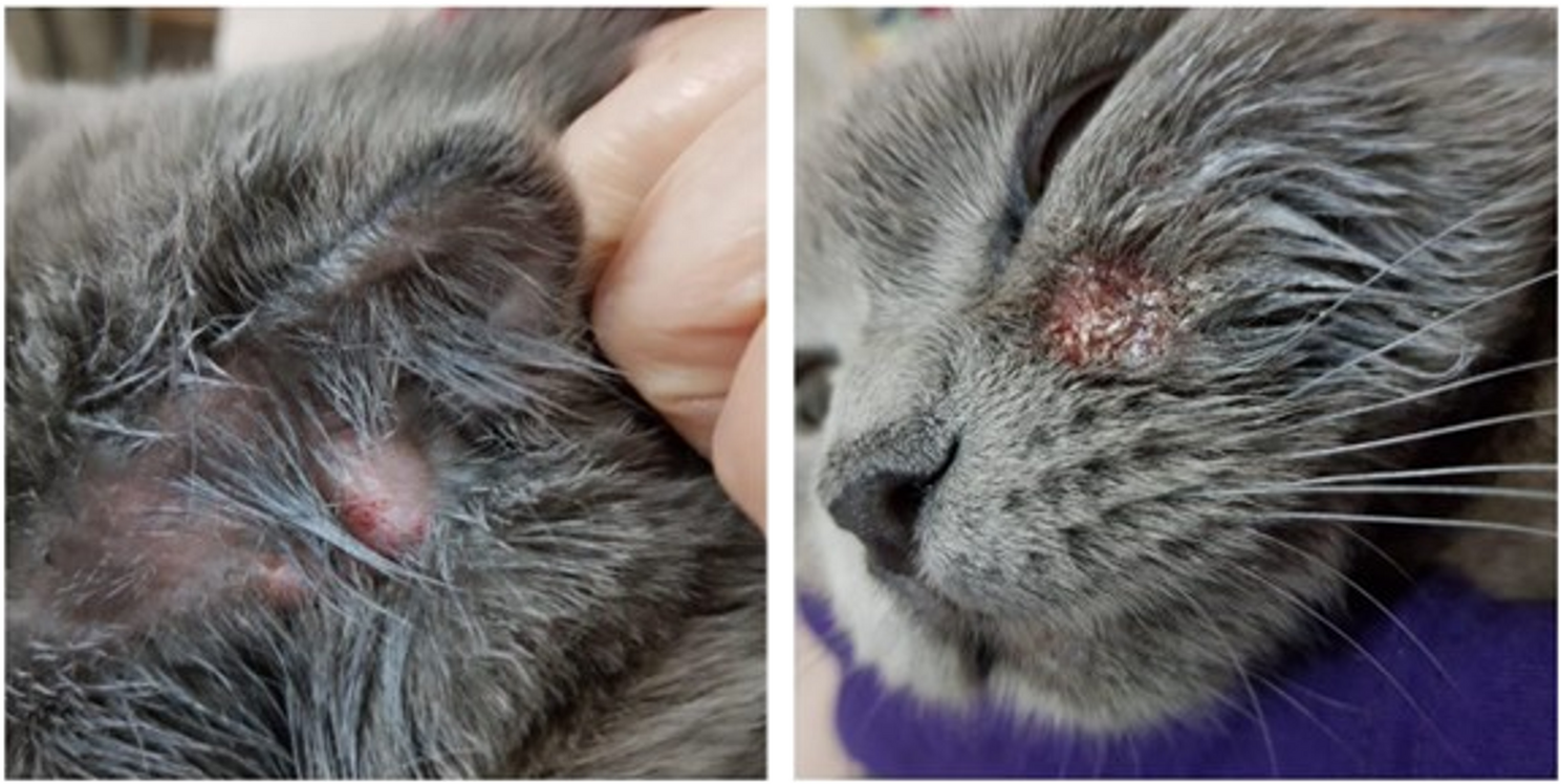
A total of 150 samples were collected from pets, including 45 dogs and 105 cats, presenting with dermatological lesions such as alopecia and peeling. These animals were brought to the Diagnostic Laboratory of the Department of Microbiology for clinical evaluation. Hair, skin scrapings, or nail samples were obtained as part of the diagnostic process for suspected dermatophytosis. No experimental intervention was performed; only diagnostic samples already submitted were used. Institutional permission and ethics approval were obtained prior to use. No animals were euthanised or sacrificed for this study. Only non-invasive specimens, including hair, skin scrapings, and nail samples, were collected for diagnostic purposes.
Fungal culture
Samples were inoculated on Sabouraud dextrose agar (Oxoid) and Dermatophyte Test Medium (DTM) (HiMedia Laboratories, Mumbai, India, Catalogue No. M188) containing cycloheximide and chloramphenicol for initial isolation. Inoculation was performed at room temperature and incubated for 3 weeks to support optimal fungal growth. Colonies observed on Sabouraud dextrose agar (SDA) were white or yellowish in colour, with a smooth, velvety, or cottony surface, and a brown to golden-yellow reverse. These macroscopic characteristics were consistent with Microsporum spp. On DTM, white aerial hyphae and a red colour change around the colony indicated the presence of dermatophytes, including Microsporum, Trichophyton, and Epidermophyton species; however, these findings are not exclusive to any particular genus. Colonies with a smooth, powdery, or granular appearance, a surface colour ranging from white to cream, and a reverse side varying from yellowish-brown to reddish-brown are features commonly associated with Trichophyton species, although similar morphologies may occasionally be observed in other dermatophyte genera as well. Therefore, while red discolouration in DTM and colony morphology are supportive in diagnosis, definitive species identification was confirmed through advanced mycological methods and sequence analysis. The macroscopic morphology of the fungal isolate was consistent with T. rubrum, as described in standard mycological literature. On Sabouraud dextrose agar, colonies initially appeared white and cottony, gradually becoming more powdery in texture with maturation. The reverse side exhibited a deep red to wine-coloured pigmentation, a hallmark feature aiding in the phenotypic identification of T. rubrum. Subsequently, the colonies were stained with lactophenol cotton blue, and the microscopic morphology was examined under a light microscope at 40× magnification [15, 16].
Molecular identification of dermatophytes
DNA used for molecular analyses, including PCR, was extracted from pure fungal cultures obtained by culturing clinical specimens (hair, skin scrapings, and nail material) collected from lesioned animals; no mixed cultures were used. For DNA extraction, 20 µl of 0.1 M dithiothreitol was added to the samples, and the procedure was performed according to the manufacturer’s instructions (Sherlock AX, A&A Biotechnology). Incubation was continued at 50 °C until all samples were lysed. The DNA obtained was stored at −20 °C until it was used in PCR analysis.
First, PCR targeting the Chitin Synthase 1 (CHS1) gene was performed on all 38 culture-positive samples using universal primers CHS1 F (5′-GAA GCC TGG AAG AAG ATT GTC G-3′) and CHS1 R (5′-CCT TGA TTT CAC CGC AGG CAC-3′), which amplify a 432 bp fragment to confirm the presence of dermatophytes [17, 18].
Second, species-specific PCR targeting the ITS region was used to confirm the presence of M. canis and T. rubrum, using respective primers and expected amplicon sizes Primer pairs for the detection of M. canis and T. rubrum were designed by targeting the internal transcribed spacer (ITS) regions of the nuclear ribosomal DNA, which are widely used in fungal diagnostics due to their ability to differentiate between genera and closely related species. The corresponding ITS sequences were retrieved from the NCBI GenBank database in FASTA format (GenBank accession numbers: T. rubrum – AF170472.1, M. canis – KY070120.1). Primer design was performed using Primer3 and NCBI Primer-BLAST tools, resulting in target amplicon lengths of approximately 557 bp for T. rubrum and 383 bp for M. canis. The candidate primers were evaluated through BLAST analysis against the NCBI nucleotide database. For T. rubrum, the forward primer (5′-CCACGATAGGGACCGACGTT-3′) and reverse primer (5′-GATTGGGGCCAGGGCG-3′) were used. For M. canis, the forward primer (5′-ATACCGTCTGAGCGAGCAAC-3′) and reverse primer (5′-TACATGGTGCGTTAGGCCAG-3′) were applied.
All primers were evaluated using Thermo Fisher’s Multiple Primer Analyzer to assess potential self- and cross-dimer formation (https://www.thermofisher.com/tr/en/home/brands/thermo-scientific/molecular-biology/molecular-biology-learning-center/molecular-biology-resource-library/thermo-scientific-web-tools/multiple-primer-analyzer.html). Melting temperatures (Tm) were calculated, and primers were selected to fall within a ± 3 °C window. Gradient PCR was performed between 60 °C and 65 °C to empirically determine the optimal annealing temperature, based on the calculated melting temperatures of the primers.
Each PCR reaction was prepared in a final volume of 50 µL, containing 25 µL of 2× PCR Master Mix (DreamTaq Hot Start Green PCR Master Mix 2X, Thermo Fisher Scientific, Waltham, MA, which includes Taq DNA polymerase, dNTPs, MgCl₂, and loading dye), 1 µL of each primer (10 µM), 2 µL of purified DNA (100 ng), and sterile ultrapure water added to complete the final volume. The PCR protocol included an initial denaturation at 96 °C for 4 min. This was followed by a stage consisting of 5 cycles of denaturation at 94 °C for 30 s, annealing at 64 °C for 60 s, and extension at 72 °C for 90 s. Subsequently, 35 cycles of standard amplification were carried out with denaturation at 94 °C for 30 s, annealing at 62 °C for 60 s, and extension at 72 °C for 90 s. The PCR was completed with a final extension step at 72 °C for 5 min [17, 18]. A 1.5% agarose gel containing 5 µg/ml ethidium bromide was prepared for visualisation and subjected to electrophoresis [17, 19, 20]. Using a 100 bp (BioTech) DNA ladder as a marker, bands of 432 bp in the CHS1 gene region, 383 bp in M. canis and 557 bp in T. rubrum were considered positive.
ITS region sequencing of the rRNA gene was used to confirm the species-level identification of selected isolates. Two T. rubrum PCR-positive isolates were selected for sequencing. Fungal isolates were sent to a commercial sequencing company, which performed DNA extraction, ITS amplification using ITS1 and ITS4 primers, and bidirectional sequencing [49]. Raw sequencing chromatograms were analysed using standard bioinformatics software to generate consensus sequences for each isolate. Consensus sequences obtained from ITS region amplification were compared with reference sequences available in the NCBI GenBank database using the BLASTn algorithm to assess species-level identification. Trichophyton rubrum (GenBank accession no. AF170472) was included as a reference sequence. Species identification was confirmed based on the highest percentage identity and high query coverage values [21, 22].
The two T. rubrum-positive isolates were further confirmed by ITS sequencing, which revealed > 99% identity to GenBank reference strains. The remaining 12 culture-positive samples could not be identified at the species level due to the absence of species-specific amplification, resulting from low DNA quality, primer mismatch, or the presence of non-target dermatophyte species.
Statistical methods
For comparisons involving categorical variables such as dermatophyte positivity by age group and species., Pearson’s chi-square test (χ²) was applied. Additionally, ANOVA was used where applicable for continuous variables. All analyses were performed using IBM SPSS Statistics 23, and p-values less than 0.01 were considered statistically significant.