
The first-ever cellular atlas of the Aedes aegypti mosquito has been created by scientists from Rockefeller University and a global team of experts.
The yellow fever mosquito is a global health threat. No other mosquito species transmits as many diseases as this one, making it one of the most dangerous animals on Earth.
Revealed on October 30, the Mosquito Cell Atlas offers cellular-level resolution of gene expression across 19 different tissues, from head to toe.
“This is a comprehensive snapshot of what every cell in the mosquito is doing as far as expressing genes. It’s a real achievement because we profiled so many different types of tissues in both males and females,” said Leslie Vosshall, who has studied this species for over two decades.
The large dataset, which profiles over 367,000 cell nuclei, is freely available to the public and scientific community.
Atlas includes both male and female mosquitoes
Before the creation of this atlas, research into the cellular biology of Aedes aegypti was fragmented and often incomplete.
Scientists were studying the mosquito’s cells, organ by organ and tissue by tissue, in separate studies.
On the other hand, prior studies disproportionately focused on the female mosquito because it requires a blood meal to reproduce and is therefore the sole vector for pathogens.
This created an “enormous bias,” leaving very little information about the male.
The researchers sought to fill this gap by creating a single, comprehensive resource that encompasses both male and female mosquitoes.
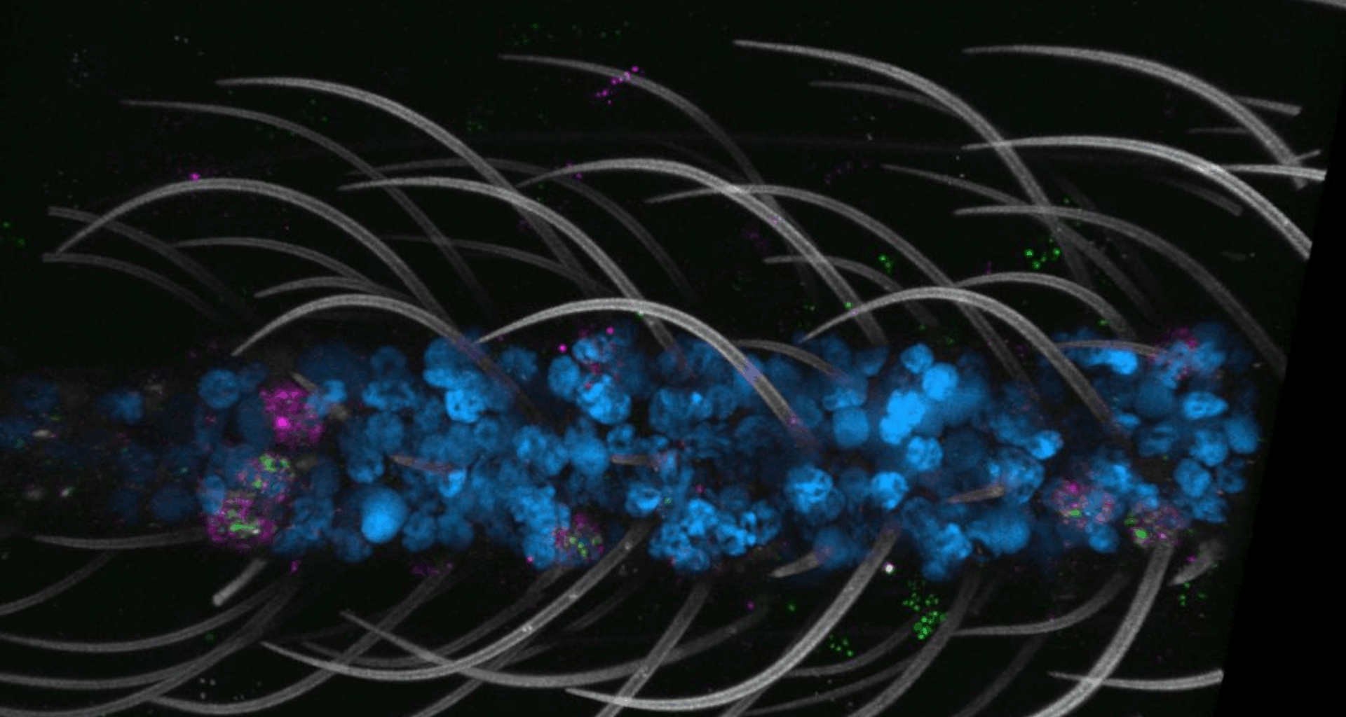
The single-nucleus RNA sequencing (snRNA-seq) technique was used to create the atlas.
The sampling included 19 types of mosquito tissues, chosen to explore five major biological functions: sensation and host-seeking behavior, viral infection, reproduction, and the central nervous system.
It successfully identified 69 distinct cell types organized into 14 major categories, with many of these cell types being novel and previously unobserved.
Cellular secrets uncovered
The atlas revealed that polymodal sensory neurons — “supercharged cells” that detect cues like temperature and taste — are far more widespread than previously known.
Although initially mapped to the antennae, the new data revealed that these multifunctional chemoreceptors are present throughout the mosquito’s body, including its nose, tongue, and legs.
“Together they enable mosquitoes to be really good at what they do—seek hosts, feed on them, and reproduce,” said Nadav Shai, senior author.
These versatile chemoreceptors enhance the mosquito’s survival, allowing the insects to detect sweetness and fresh water.
The atlas also showcased brain rewiring after a blood meal.
After a female mosquito takes a blood meal, her interest in hosts (humans) is turned off to focus on developing eggs. The atlas showed dramatic changes in gene expression in the brain during this time.
A striking and unexpected finding was that the shift in a female mosquito — i.e., losing the urge to bite after a meal — is primarily driven by changes in the glia.
New atlas: A global resource
Although glia are support cells that account for less than 10% of the brain’s cells, the researchers observed that these cells are “completely rewired” during this period.
Another surprising finding from the atlas was the limited sexual dimorphism at the cellular level despite the many documented differences in morphology and behavior between male and female mosquitoes.
The atlas showcased that the cellular makeup of male and female mosquitoes is largely identical.
The only major exceptions were the reproductive organs and small clusters of sex-specific cells.
The newly published atlas will function as a global resource to propel future mosquito research, leading to new discoveries.
The study was published in the journal Cell on October 30.