
Maintenance of P. falciparum asexual-blood stages and induction of gametocytogenesis
Plasmodium falciparum strains 3D7, NF54-E (MRA-1000), Dd2, and Dd2-GNF156, which were obtained from Bei Resources (USA), were maintained in culture as previously described [29] in fresh O-positive human red blood cells suspended at 4% (v/v) haematocrit in complete RPMI 1640 medium (500 mL RPMI 1640 (Sigma, Munich, Germany) supplemented with 25 mM HEPES, 10% Albumax I (Gibco, Waltham, MA, USA), 1X hypoxanthine (Gibco, Waltham, MA, USA) and 50 mg/mL gentamicin (Sigma-Aldrich, Munich, Germany) and incubated at 37 °C in a humidified atmosphere containing 5% CO2. The medium was renewed daily to propagate the culture. Parasite growth was monitored by microscopic examination of Giemsa-stained thin blood smears using immersion oil and synchronized at the ring stage by serial treatment with 5% D-sorbitol two days before each experiment [30]. Gametocytogenesis was induced from synchronized asexual P. falciparum parasite strain NF54-E (MRA-1000) and cultured as previously established [31].
Inhibition of P. falciparum asexual blood stages
Subfractions and the isolated compound were evaluated for their inhibitory potential on chloroquine-sensitive (Pf3D7; multi-resistant (PfDd2) and mutant-resistant (PfDd2-GNF156) strains of P. falciparum using PfLDH assay as previously described [32, 33]. In brief, 10 µl of subfractions and the isolated compound and 90 μL of synchronized parasites (ring stages) at 2% parasitaemia and 1% haematocrit were dispensed in each well of a microtiter plate. The test concentrations ranged from 0.04 to 25 μg/mL, and the final concentration of DMSO did not exceed 0.5%, which was found to be non-toxic for the parasite (negative control). Standard drugs were screened at the highest concentration of 1 µM. Plates were then incubated at 37 °C under a 5% CO2 atmosphere for 72 h. Afterwards, 20 µl of each well was transferred to a new plate containing 100 µl of Malstat solution [55 mM Tris, 0.22 M L-lactic acid, 0.17 mM acetylpyridine adenine dinucleotide [APAD; Sigma-Aldrich], 0.2% [v/v] Triton X-100(Sigma-Aldrich), pH 9.0] and 25 µl NBT/PES solution (1.96 mM nitrotetrazolium blue chloride, 0.24 mM phenazine ethosulphate, Sigma-Aldrich, Germany). Plates were left to develop in the dark for 30 min at room temperature before recording the absorbance at 650 nm using a microtiter plate reader (TECAN Infinite M200, USA). The data analysis was performed with GraphPad Prism 5.0, and dose–response curves were drawn to determine the median inhibitory concentrations (IC50s). The degree of resistance (resistance index; RI) was determined by comparing the antiplasmodial activity of the test sample on chloroquine-susceptible vs multidrug-resistant strains of P. falciparum using the following formula: RI: (IC50 multidrug-resistant strain) / IC50 (chloroquine-sensitive strain). The experiments were carried out three times, and the IC50 value was calculated as the mean of the three measurements taken from these trials.
Inhibition of P. falciparum late-stage IV/V gametocyte viability
To evaluate the impact of the isolated compound and subfractions on late-stage NF54-E (MRA-1000) gametocytes (> 90% stage IV/V, 2% gametocytaemia), the experimental method previously reported [31] was carried out using a P. falciparum lactate dehydrogenase (PfLDH)-based assay following 48 h of drug treatment. Dual point inhibition (at 1 and 5 μg/mL, n = 1, in technical triplicates) was used for subfractions and the isolated compound, while methylene blue was used as a positive control in this experiment. Microsoft Excel was used to calculate the percentage of gametocyte inhibition.
Haemolysis assay
A haemolysis assay was performed as previously described [34] against normal O+ human red blood cells to assess the haemolytic effect of the test subfractions and eschweilenol C at concentrations ranging from 100 to 0.16 µg/mL. Briefly, 250 µl of O+ human red blood cells at 2% haematocrit were mixed with 250 µl of the test sample at a concentration ranging from 100 to 0.16 µg/mL in an Eppendorf tube, followed by incubation for 2 h at 37 °C. Triton X-100 0.1% (v/v) and phosphate buffer saline (PBS), which induced 100% and 0% haemolysis in RBCs, were used as positive and negative controls, respectively. After 2 h of incubation at 37 °C, the preparation composed of lysed red blood cells and free haemoglobin was centrifuged at 1500 rpm for 5 min, and the supernatant was separated. Next, the amount of haemoglobin released in each supernatant was quantified spectrophotometrically at 540 nm using a microtiter plate reader (TECAN Infinite M200, USA) and compared to the negative control. The haemolysis percentage was calculated as follows:
$$\text{\% Haemolysis }=\frac{\text{Absorbance\, of \,sample }540\text{ nm }-\text{Absorbance\, of\, blank\, sample }540\text{nm }}{\text{Absorbance\, of\, positive\, control }540\text{ nm}}\times 100$$
Assessment of selectivity on normal cells
A Resazurin based-assay was used to evaluate the in vitro cytotoxicity of active subfractions and eschweilenol C as described [35] against Vero ATCC CRL 1586 (African green monkey kidney) and Raw 264.7 (macrophage) cells. These cells were cultured in a T-25 cm2 flask containing DMEM (Sigma-Aldrich, Munich, Germany) supplemented with 2 mM L-glutamine (Sigma-Aldrich, Munich, Germany) and 10% fetal bovine serum (ATCC), which were maintained at 37 °C in a humidified incubator containing 5% CO2. For each experiment, 80–90% confluent cell culture was trypsinized, counted using a Neubauer counting chamber and seeded onto 96-well flat-bottomed plates treated for cell culture (Costar, USA) at a cell density of 104 cells/well and allowed to adhere overnight. Cells were then treated with serially diluted concentrations of active inhibitors for 48 h. In the assay plates, Podophyllotoxin was included at concentrations ranging from 100 µM to 0.16 µM as positive control while a 0.5% DMSO solution, which demonstrated 100% cell viability, served as the negative control. After incubation, 10 µl of resazurin solution (0.15 mg/mL prepared in PBS) was added to each well, and plates were re-incubated for 4 h to allow the conversion of non-toxic cell-permeable resazurin (weakly fluorescent blue dye) to the highly fluorescent red dye resorufin by viable cells. The level of fluorescence that positively correlates with cell viability was measured using an Infinite M200 plate reader (Tecan, USA) at excitation and emission wavelengths of 530 nm and 590 nm, respectively. The inhibitory percentage of cell proliferation was calculated from the absorbance values of untreated and treated cells. Sigmoidal concentration–response curves were plotted using GraphPad Prism (v5), from which the half-maximal cytotoxic concentration (CC50) values were determined. Selectivity indices (SI) [SI = CC50/IC50] of test samples were determined based on their antiplasmodial activity (IC50) and cytotoxicity (CC50 on Vero and Raw cells). The experiments were conducted in triplicate, and the CC50 value was reported as the average of the three measurements obtained from the three experiments.
Haemozoin inhibition assay
The ability of active antiplasmodial subfractions and the isolated compound to inhibit the haemozoin (β-haematin) produced by P. falciparum in cultures was assessed by using drug concentrations at their IC50 (Ti030.54 µg/mL, Ti040.49 µg/mL, and eschweilenol C490.74 µM) upon incubation for 72 h (assay time) [36]. Artemisinin and chloroquine were used as positive controls. After the incubation time had elapsed, cultures were transferred in an Eppendorf tube and centrifuged for 7 min at 1300 rpm to dispose of the culture medium. The infected erythrocyte pellet, composed of membrane debris and β-haematin, was subjected to lysis by 0.01% saponin at 25 °C for 10 min to release the parasites from red blood cells. The released parasites were further washed thrice with PBS for 5 min at 1500 rpm, re-suspended in 2.5% sodium dodecyl sulfate buffer solution (SDS in PBS), and subjected to spin at 20,000 g for 1 h. The supernatant was disposed of, and the insoluble haemozoin pellet was washed in 2.5% SDS in PBS and then dissolved in 20 mM NaOH. Next, the absorbance of the preparation was read at 405 nm, and a standard curve was generated to determine the quantity of β-haematin, from which the amount of haemozoin crystals was deduced. All assays were performed in triplicate.
Time-kill kinetics
The kinetics of growth of P. falciparum multidrug-resistant strain (Dd2) was monitored upon treatment with the most promising antiplasmodial samples viz. Subfractions Ti03, Ti04, and eschweilenol C according to a previously described procedure [37]. Briefly, a preparation containing an asynchronous parasite blood culture with test samples (Ti03, Ti04, and eschweilenol C) was assessed using a PfLDH-based method to yield corresponding IC50 values. Three exposure times were employed for each test sample, viz.72 (standard assay time), 48, and 24 h. The IC50 ratios, normalized to the 72 h exposure, were subsequently used to classify test samples (Ti03, Ti04, and eschweilenol C) as fast or slow-acting.
Plasmodium falciparum asexual-blood stage-specific analysis and post-drug effect of the most promising antiplasmodial hits
The rationale for evaluating actives against the three developmental stages of the malaria parasite (rings, trophozoites, and schizonts) stems from the intricate life cycle of P. falciparum and the specific biological processes that characterize each stage. Each developmental phase exhibits distinct metabolic demands and vulnerabilities, positioning them as crucial targets for therapeutic intervention. To achieve that, highly synchronized resistant strain of P. falciparum (Dd2) rings, trophozoites, and schizonts stages (2% parasitaemia and 1% haematocrit) were treated with Ti03, Ti04 and eschweilenol C at their 99% inhibitory concentration (IC99) for 48 h (rings), 24 h (trophozoites) and 12 h (schizonts) under standard culture conditions. Giemsa-stained thin smears were prepared at different time intervals (0, 6, 24, and 48 h for rings, 0, 6, 18, and 24 h for trophozoites, and 12 h for schizonts) for microscopic examinations of the intraerythrocytic maturation of the parasite and to determine the effects on the parasite growth [38, 39] and the stage-to-stage transition. After incubation, test samples were removed by three-time media wash, and the cultures were re-suspended in a complete culture medium and maintained under standard culture conditions for 48 h in rings, 24 h in trophozoites, and schizonts. Next, thin smears were prepared from control and treated wells and processed for Giemsa staining. The percentage of stage-specific inhibition by Ti03, Ti04, and eschweilenol was compared to the corresponding drug-free control by counting 3000 cells (10–12 fields) for each parasite stage. Parasitized and non-parasitized cells were counted by microscopy to determine stage-specific growth inhibition, cytostatic or cytocidal effect, and growth suppression following post-sample exposure on P. falciparum Dd2. The parasites with pyknotic morphology were considered nonviable cells (death parasites).
In silico molecular docking and ADME studies of eschweilenol
In silico molecular docking studies were performed to support the pharmacodynamics investigations and inhibition of haemozoin formation. Research has been concentrated on P. falciparum M1 alanyl aminopeptidase (PfM1AAP) and P. falciparum M17 leucyl aminopeptidase (PfM17LAP). These enzymes play a critical role in the final stages of haemoglobin degradation, producing vital amino acids that support parasite growth. Consequently, they present promising opportunities for the development of novel anti-malarial drugs.
Structure drawing, cleaning, and optimization
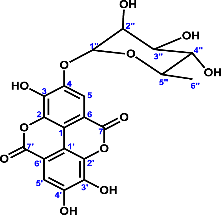
ChemDraw Professional version 15.0 software (PerkinElmer Informatics, Waltham, MA, USA) was used to clean the drawing and geometry of the antiplasmodial compound eschweilenol C. The two-dimensional (2D) structures were transformed into three-dimensional (3D) structures using Chem3D version 15.0. Afterwards, the energy of the 3D structure was minimized using a molecular mechanics-2 (MM2) force field.
Molecular docking and binding energy studies
The docking analysis of selected targets (P. falciparum M1 alanyl aminopeptidase (3EBG), P. falciparum M17 leucyl aminopeptidase (3KQX)) and the ligand eschweilenol C was accomplished by employing AutoDock Vina v1.5.7 (Molecular Graphics Lab at The Scripps Research Institute, La Jolla, CA92037, USA). 3D crystallographic structures of the target enzymes were retrieved from the RCSB Protein Data Bank. An expanded version of the PDB format was employed for the coordinate files, including atomic partial charges and PDBQT files. The software automatically transformed the PDB files to PDBQT, which was then used in the docking process. To obtain the appropriate tautomeric and ionized states, polar hydrogen atoms were added to the target proteins for amino acid residues. The native ligand of the co-crystallized complex was first removed and re-docked to its matching binding pocket using AutoDock Vina v1.5.7 to standardize the software. The ultimate binding/docking energy was determined by averaging the three times the ligands and receptors were docked. For each target protein, the binding poses with the lowest docking energy served as the ultimate model for the post-docking study. This analysis was done using PyMOL (The PyMOL Molecular Graphics System, Version 1.5.0.4 Schrodinger, LLC, USA) and UCSF Chimera v1.5.3 (NCRR, University of California, San Francisco, supported by NIH P41 RR001081).
Virtual prediction of pharmacokinetics properties
The ADMET (absorption, distribution, metabolism, excretion, and toxicity) properties of drugs are fundamental in assessing their therapeutic potential and safety profile across all human diseases [40]. In this context, the ADME parameters and Lipinski rule of eschweilenol C were analysed using a free online tool, ADMETlab 2.0 (https://admetmesh.scbdd.com/). All the studied substances were transformed into their standard Simplified Molecular Input Line Entry System (SMILES) format and then uploaded to ADMETlab 2.0 for analysis [41]. This web server provides the users easy access to the comprehensive, accurate, and efficient prediction of the ADMET profiles for chemicals, including absorption, distribution, metabolism, excretion, toxicity properties, and some essential physicochemical and medicinal chemistry properties. Significant chemical descriptors, such as polar surface area (PSA), play a crucial role in fraction absorption, and low molecular weight (MW) is essential for oral absorption. Therefore, the bioavailability of the eschweilenol C was assessed using TPSA analysis.