
By John Lewis of 
The rare and potentially transmissable condition is caused by medical procedures involving exposure to pathological amyloid-beta protein ”seeds”, which travel to the brain.
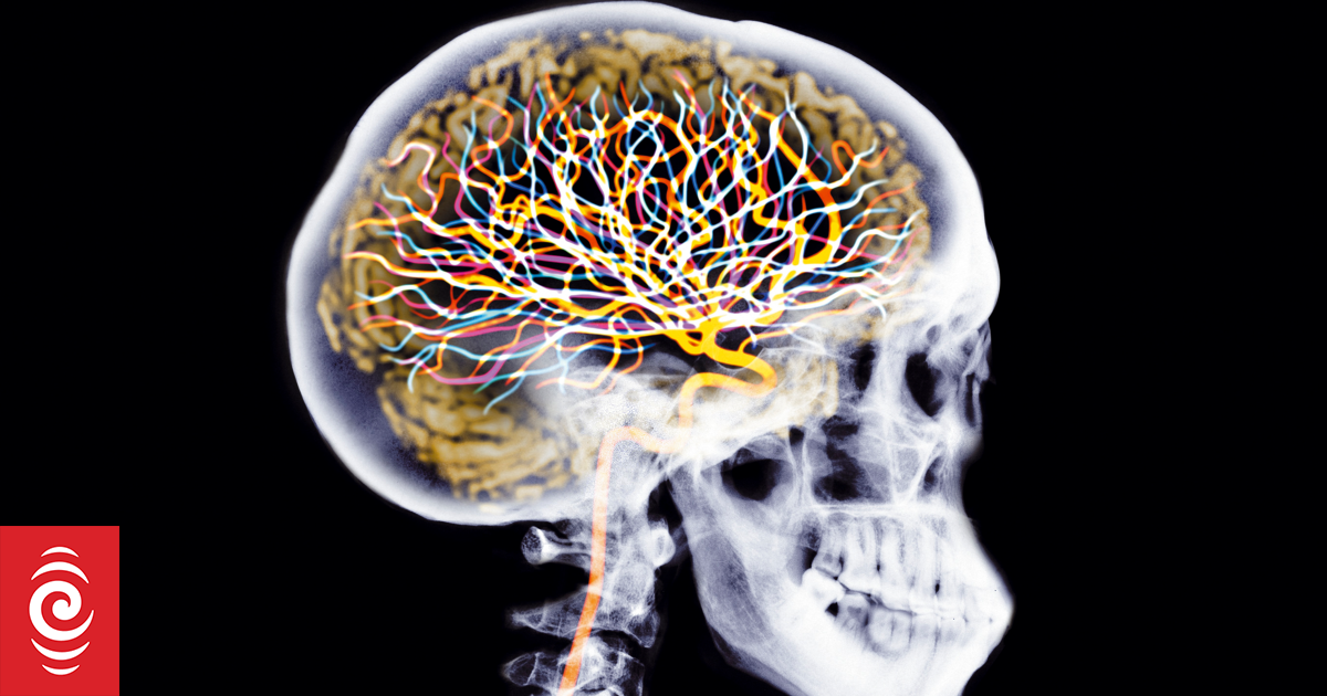
Photo: NICK VEASEY/ SCIENCE PHOTO LIBRARY / AFP
A patient given a dural graft from a cadaver at Dunedin Hospital as a child in the 1980s has recently been diagnosed as having iatrogenic cerebral amyloid angiopathy (iCAA).
The rare and potentially transmissable condition is caused by medical procedures involving exposure to pathological amyloid-beta protein ”seeds”, which travel to the brain.
Research published in the New Zealand Medical Journal by Chuen Siang Low, Sarah Buchanan, Nicholas Cutfield, Sophie Parker and Robin Fox showed the patient was born in the early 1980s with aplasia cutis congenita – the localised absence of skin, most commonly on the scalp.
It exposed the dura – the underlying outermost membrane which protects the brain.
”In the first weeks of life, the dura deteriorated, necessitating surgical procedures to resect non-viable herniated brain tissue and to close the dural defect.
”The dura was repaired with lyophilised cadaveric dura.”
While the surgery left him with weakness, or partial paralysis, on the left side of his body, as well as mild intellectual disability and focal epilepsy, he had been clinically stable for many years and had been living independently and working full-time.
However, now at age 43, the man with a long-standing history of focal epilepsy presented with increasing seizure frequency, cognitive decline and behavioural changes.
Testing of his cerebrospinal fluid results, as well as magnetic resonance imaging of his brain, found his condition was likely to be the result of iCAA.
The researchers believe it is the first reported case of iCAA in New Zealand.
”On assessment, he was disorientated and inattentive, anxious and expressing paranoid thoughts.”
The weakness on his left side was also worse than his baseline, and there were frequent bouts of left-sided face, arm and leg twitching, suggestive of focal seizures.
Brain MRI scans showed multiple white matter changes, often associated with age, chronic hypertension, or small vessel disease.
They also showed numerous bilateral micro-haemorrhages.
Given the appearance of cortical micro-bleeds, and the history of exposure to cadaveric dural tissue, the researchers tested for iCAA.
The tests found decreased cerebrospinal fluid amyloid-beta levels, which was indicative of amyloid deposition in the central nervous system, and it supported their diagnosis of probable iCAA.
”The patient was discharged from hospital on an adjusted anti-seizure medication regimen with acceptable seizure control, and improvements in paranoia.
”Unfortunately, there has not been a significant cognitive or functional improvement, and he requires hospital-level care.”
The research said the patient and his family had been counselled about the risk of future intracranial bleeding.
There have been about 50 cases of iCAA reported in literature, and the onset of clinical symptoms can be reported between 25 and 46 years after exposure to cadaveric dural tissue.
”Cadaveric lyophilised dural grafts are one of the commonest reported causes of iCAA, and were used in neurosurgical procedures in New Zealand in the 1980s,” the research said.
”Usage ceased when they were recognised as a cause of iatrogenic Creutzfeldt-Jakob disease, the best-known example of seeding of abnormal proteins causing a neurodegenerative condition.”
It was unknown how many patients in New Zealand received cadaveric dural grafts, because no records were kept.
The researchers said this case highlighted the importance of considering iCAA in younger patients with a history of dural graft use.
– This story first appeared in The Otago Daily Times