
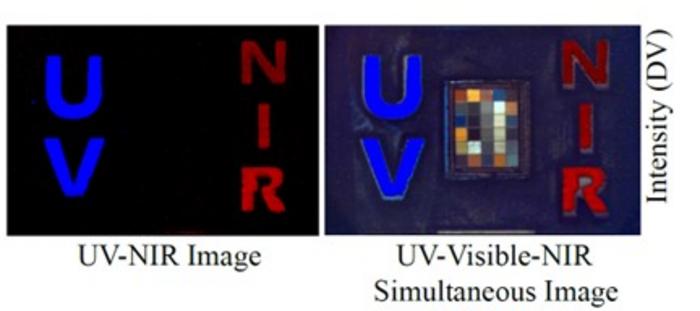
 Shown are single exposures capturing deep UV fluorescence (tryptophan), visible color (Macbeth chart) and NIR fluorescence (ICG).
Credit©
Viktor Gruev, University of Illinois at Urbana-Champaign
Researchers have created a camera inspired by mantis shrimp vision that sees UV, infrared, and visible light on a single chip. In tests with breast cancer patients, the device was 97% sensitive in identifying cancerous lymph nodes during surgery, potentially allowing for more precise, less invasive procedures
Surgeons often face a difficult choice during cancer surgery: which lymph nodes to remove. Removing too many can lead to lifelong complications like lymphedema, while removing too few risks leaving cancer behind. A new study in Optica reveals a compact, single-chip camera that could solve this dilemma.
Inspired by the extraordinary eyes of the mantis shrimp, researchers at the University of Illinois at Urbana-Champaign have developed a tool that helps surgeons locate and assess cancerous lymph nodes in real-time.
Bio-inspired multi-spectrum vision: Detecting cancerous lymph nodes
The mantis shrimp is famous for its complex eyes, which contain stacked rows of photoreceptors capable of seeing ultraviolet (UV), visible, and near-infrared (NIR) light simultaneously. Human eyes—and most standard cameras—cannot do this without bulky extra equipment.
The researchers replicated this biology on a single chip:
NIR channel:
Detects a common dye called indocyanine green (ICG) to map exactly where lymph is draining from a tumour.
UV channel:
Captures the tissue’s natural fluorescence (tryptophan), which changes when cancer is present. This allows for “label-free” assessment—detecting cancer without needing a specific chemical marker.
Visible channel:
Provides the standard colour view so surgeons can see the anatomical context of the surgery.
Precision surgery without the wait
Current methods for checking if cancer has spread to a lymph node often involve sending tissue to a lab while the patient is still on the operating table, which can take 20 to 30 minutes.
The mantis-shrimp camera provides this information instantly. Because all three light signals are captured by the same chip at the same time, the images are perfectly aligned. This “all-in-one” view allows a surgeon to see exactly where a node is (via NIR) and immediately judge if it looks suspicious (via UV).
High accuracy in human testing
The team tested the camera on 94 lymph nodes from 33 breast cancer patients. When compared to traditional pathology results, the camera’s UV readout achieved 97% sensitivity and 89% specificity in identifying metastatic cancer.
While these results are incredibly promising, the researchers emphasise that the camera is intended to be a decision-support tool, helping surgeons make more informed choices rather than replacing their expert judgment. The next steps involve refining the hardware for sterile operating rooms and testing the system on other types of cancer where lymph node status is critical.