
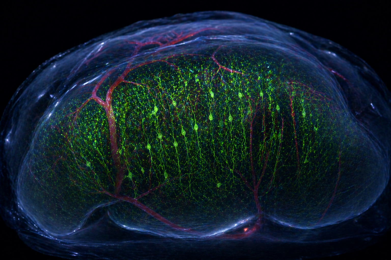
 Fluorescence imaging of the penultimate layer of the cerebral cortex of the mouse brain before (left) and after (right) the use of the researchers’ new SeeDB-Live clearing agent. Credit: Shigenori Inagaki & Takeshi Imai/Kyushu University
Researchers have figured out how to temporarily look straight through a living brain tissue with minimal invasiveness and interference in its processes.
Japanese researchers at Kyushu University discovered a chemical key that temporarily creates a highly transparent window into a living brain, revealing the flashes of individual neurons as they fire deep within the cortex.
By harnessing a common protein naturally found in blood, the method allows for much deeper imaging in acute brain slices and living mice, all while the animal’s brain remains fully functional, healthy, and actively thinking.
Marbles in Oil
Imagine dropping a handful of glass marbles into a bowl of water. You can still see the marbles because light bends slightly as it passes from the water into the glass. If you drop those same marbles into a bowl of heavy oil, however, they seem to vanish. That’s because the glass and the oil share the exact same refractive index, allowing light to travel straight through them without scattering.
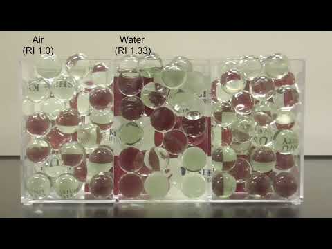
Marble balls refractive index experiment. Credit: Kyushu University.
Brain tissue is an optical mess. Water, lipids, and tangled cellular membranes all bend light in different directions. If researchers could adjust the fluid surrounding these cells to match the refractive index of the cells themselves, the brain would turn transparent.
Takeshi Imai, a biochemist and professor at Kyushu University’s Faculty of Medical Sciences, has chased this optical illusion for years. He previously developed chemicals that successfully turned dead, preserved tissue transparent (SeeDB & SeeDB2). Moving that technique into living, breathing animals was a monumental hurdle.
“When we first made postmortem specimen mouse brains transparent more than a decade ago, people often asked when we would be able to do it with a living mouse,” Imai said in a statement.
“We have finally managed it. Since we can now observe the brain without affecting its functions, I hope researchers will make use of this in a wide variety of studies.”
×
Thank you! One more thing…
Please check your inbox and confirm your subscription.
A Desperate Midnight Gamble
Finding the exact optical sweet spot—a refractive index between 1.36 and 1.37—was only half the battle. The team needed a chemical that would not kill the brain cells it illuminated.
Living cells are like delicate water balloons. Exposing them to a dense, sugary solution, and osmosis will violently suck the moisture out of them, causing the cells to dehydrate and die. The researchers realized they needed massive, spherical molecules. Large molecules generate much less osmotic pressure, meaning they could change how the tissue handles light without crushing the delicate cells.
Shigenori Inagaki, an assistant professor working with Imai, took on the grueling task of testing nearly 100 different polymers.
“I started working on this around 2021, and within a few months, I concluded that spherical polymers seemed to be the way to go,” Inagaki recalled. “However, the candidate molecules were adversely affecting cell function, and I was stuck for about a year. When I was working alone in the lab late at night and there was no one around, I tried a highly pure, expensive albumin reagent—and it worked.”
Albumin is a highly soluble blood-serum protein; the team used Bovine Serum Albumin (BSA), a standard lab reagent. Acting entirely on a hunch, Inagaki applied BSA to his samples.
“I tested it three or four times before I believed it,” Inagaki says. “Of all things, we never expected it would come down to this.”
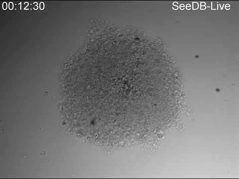
Three-dimensional cell aggregates (spheroids) become transparent within 15 minutes when immersed in SeeDB-Live.
Seeing Deep Brain Live
The team named their new solution SeeDB-Live. It operates with startling efficiency. When scientists immerse a slice of living mouse brain into the solution, the opaque gray matter turns transparent within 30 minutes to one hour.
“During the development of SeeDB-Live, we found that neurons are extremely sensitive to ion concentrations, and it took us enormous effort to get the formulation right,” Inagaki added. “Thanks to that fortunate night alone in the lab, I helped myself to an expensive, high-purity BSA I wouldn’t normally dare use.”
By pairing SeeDB-Live with fluorescent calcium indicators—chemical tags that physically light up when a nerve fires—the team achieved something unprecedented. They peered all the way down into the fifth layer of the cerebral cortex. Layer 5 contains large projection neurons that help send cortical output to other brain regions and the body.

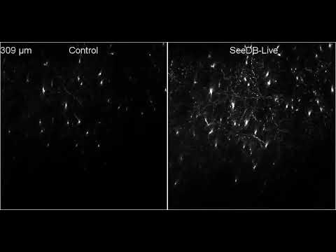
Time-lapse two-photon imaging of the calcium indicator, GCaMP6f, in an acute slice of a 11-day-old mouse olfactory bulb (depth, 150 μm). The fluorescence gradually increased, enabling calcium signal recordings from neurons at tissue depths that are typically difficult to access.
In living mouse brains, fluorescence signals from deep neurons became about three times brighter. The improved brightness made deep neuronal structures and activity easier to record.
“This is the first time tissue clearing has been achieved without altering its biology,” says Imai.
Unlike previous harsh chemicals, SeeDB-Live does not permanently alter the animal. Within a few hours, the bodily fluids naturally wash the albumin out of the extracellular space. The transparent brain simply fogs back up, returning to its natural state. This gentle reversal allowed the team to image the exact same mice repeatedly over four months, tracking shifting neural activity over long periods.

A mouse brain slice (5 days old, 300 μm thick) was cleared with SeeDB-Live.
The Elegant Logic of Evolution
The secret to this radical new imaging technique was hiding in plain sight, flowing through the veins of the very animals the scientists were studying.
“Albumin is abundant in blood and highly soluble, which makes it well-suited for clearing,” notes Imai. “It was an accidental discovery, but looking back, it feels almost natural. What evolution has shaped over millions of years is truly impressive.”
Biologists now possess a tool to observe exactly how healthy brains process information, or how diseases like Alzheimer’s disrupt those fragile networks. Furthermore, the reagent could allow pharmaceutical researchers to look inside artificially grown brain organoids, observing exactly how experimental drugs alter living neural circuits.
“SeeDB-Live can pave the way for deep-tissue live imaging, both ex vivo and in vivo,” says Inagaki.
Fluorescence images of the cerebral cortex of a Thy1-YFP-H transgenic mouse, before and after clearing with SeeDB-Live.
Challenges remain. Currently, biologists must surgically create a small window in the mouse’s skull to apply the solution directly to the brain surface.
“I feel we have not yet fully materialized its potential,” Inagaki says. The team is looking toward future experiments focused on less invasive delivery methods.
For Imai, gazing into a living, functioning mind marks the end of a long, frustrating road.
“That question came to me about a hundred times, and each time I answered ‘impossible,’” Imai says. “But 10 years later, here we are. When something seems unachievable, if you keep thinking about it, you may eventually find a way.”
The study was published in the journal Nature Methods.